 Movie
Movie Controller

[English] 日本語
 Yorodumi
- PDB-4utq: A structural model of the active ribosome-bound membrane protein ... -
+ Open data

- Basic information
| Entry | Database: PDB / ID: 4utq | ||||||
|---|---|---|---|---|---|---|---|
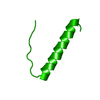
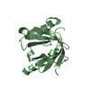
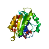
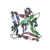
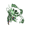
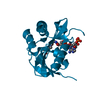
| Title | A structural model of the active ribosome-bound membrane protein insertase YidC | ||||||
 Components Components |
| ||||||
 Keywords Keywords | PROTEIN TRANSPORT / PROTEIN TRANSLOCATION / BIOINFORMATICS / MD SIMULATION / MEMBRANE | ||||||
| Function / homology |  Function and homology information Function and homology informationmembrane insertase activity / cell envelope Sec protein transport complex / protein transport by the Sec complex / proton motive force-driven plasma membrane ATP synthesis / proton motive force-driven ATP synthesis / proton-transporting two-sector ATPase complex, proton-transporting domain / protein insertion into membrane / proton-transporting ATPase activity, rotational mechanism / proton-transporting ATP synthase complex / proton-transporting ATP synthase activity, rotational mechanism ...membrane insertase activity / cell envelope Sec protein transport complex / protein transport by the Sec complex / proton motive force-driven plasma membrane ATP synthesis / proton motive force-driven ATP synthesis / proton-transporting two-sector ATPase complex, proton-transporting domain / protein insertion into membrane / proton-transporting ATPase activity, rotational mechanism / proton-transporting ATP synthase complex / proton-transporting ATP synthase activity, rotational mechanism / protein folding / protein-containing complex assembly / lipid binding / membrane / plasma membrane Similarity search - Function | ||||||
| Biological species |  | ||||||
| Method | ELECTRON MICROSCOPY / single particle reconstruction / cryo EM / Resolution: 8 Å | ||||||
 Authors Authors | Wickles, S. / Singharoy, A. / Andreani, J. / Seemayer, S. / Bischoff, L. / Berninghausen, O. / Soeding, J. / Schulten, K. / vanderSluis, E.O. / Beckmann, R. | ||||||
 Citation Citation | Journal: Elife / Year: 2014 Title: A structural model of the active ribosome-bound membrane protein insertase YidC. Authors: Stephan Wickles / Abhishek Singharoy / Jessica Andreani / Stefan Seemayer / Lukas Bischoff / Otto Berninghausen / Johannes Soeding / Klaus Schulten / Eli O van der Sluis / Roland Beckmann /   Abstract: The integration of most membrane proteins into the cytoplasmic membrane of bacteria occurs co-translationally. The universally conserved YidC protein mediates this process either individually as a ...The integration of most membrane proteins into the cytoplasmic membrane of bacteria occurs co-translationally. The universally conserved YidC protein mediates this process either individually as a membrane protein insertase, or in concert with the SecY complex. Here, we present a structural model of YidC based on evolutionary co-variation analysis, lipid-versus-protein-exposure and molecular dynamics simulations. The model suggests a distinctive arrangement of the conserved five transmembrane domains and a helical hairpin between transmembrane segment 2 (TM2) and TM3 on the cytoplasmic membrane surface. The model was used for docking into a cryo-electron microscopy reconstruction of a translating YidC-ribosome complex carrying the YidC substrate FOc. This structure reveals how a single copy of YidC interacts with the ribosome at the ribosomal tunnel exit and identifies a site for membrane protein insertion at the YidC protein-lipid interface. Together, these data suggest a mechanism for the co-translational mode of YidC-mediated membrane protein insertion. | ||||||
| History |
|
- Structure visualization
| Movie |
Movie viewer |
|---|---|
| Structure viewer | Molecule: MolmilJmol/JSmol |
- Downloads & links
-Download
| PDBx/mmCIF format | 4utq.cif.gz | 37.9 KB | Display | PDBx/mmCIF format |
|---|---|---|---|---|
| PDB format | pdb4utq.ent.gz | 16.5 KB | Display | PDB format |
| PDBx/mmJSON format | 4utq.json.gz | Tree view | PDBx/mmJSON format | |
| Others |  Other downloads Other downloads |
-Validation report
| Arichive directory | https://data.pdbj.org/pub/pdb/validation_reports/ut/4utqftp://data.pdbj.org/pub/pdb/validation_reports/ut/4utq | HTTPS FTP |
|---|
-Related structure data
| Related structure data | 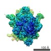 2705MC M: map data used to model this data C: citing same article ( |
|---|---|
| Similar structure data |
-Links
 PDBj
PDBj


- Assembly
| Deposited unit | 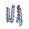
|
|---|---|
| 1 |
|
-Components
| #1: Protein | Mass: 61576.508 Da / Num. of mol.: 1 / Source method: isolated from a natural source / Source: (natural) |
|---|---|
| #2: Protein | Mass: 8259.064 Da / Num. of mol.: 1 / Fragment: MEMBRANE DOMAIN / Source method: isolated from a natural source / Source: (natural) |
-Experimental details
-Experiment
| Experiment | Method: ELECTRON MICROSCOPY |
|---|---|
| EM experiment | Aggregation state: PARTICLE / 3D reconstruction method: single particle reconstruction |
- Sample preparation
| Component | Name: MONOMERIC YIDC BOUND TO RIBOSOME NASCENT CHAIN COMPLEX(F0C) Type: RIBOSOME |
|---|---|
| Specimen | Embedding applied: NO / Shadowing applied: NO / Staining applied: NO / Vitrification applied: YES |
| Specimen support | Details: CARBON |
| Vitrification | Instrument: FEI VITROBOT MARK IV / Cryogen name: ETHANE Details: VITRIFICATION 1 -- CRYOGEN- ETHANE, INSTRUMENT- FEI VITROBOT MARK IV, |
- Electron microscopy imaging
| Experimental equipment |  Model: Titan Krios / Image courtesy: FEI Company |
|---|---|
| Microscopy | Model: FEI TITAN KRIOS / Date: Jun 1, 2013 |
| Electron gun | Electron source:  FIELD EMISSION GUN / Accelerating voltage: 200 kV / Illumination mode: SPOT SCAN FIELD EMISSION GUN / Accelerating voltage: 200 kV / Illumination mode: SPOT SCAN |
| Electron lens | Mode: BRIGHT FIELD / Nominal defocus max: 3500 nm / Nominal defocus min: 1300 nm |
| Image recording | Electron dose: 20 e/Å2 / Film or detector model: TVIPS TEMCAM-F416 (4k x 4k) |
| Image scans | Num. digital images: 2000 |
- Processing
| EM software | Name: SPIDER / Category: 3D reconstruction | ||||||||||||
|---|---|---|---|---|---|---|---|---|---|---|---|---|---|
| CTF correction | Details: DEFOCUS GROUP | ||||||||||||
| Symmetry | Point symmetry: C1 (asymmetric) | ||||||||||||
| 3D reconstruction | Resolution: 8 Å / Num. of particles: 58960 / Nominal pixel size: 1.035 Å Details: ONLY THE MEMBRANE PART WAS MODELED SUBMISSION BASED ON EXPERIMENTAL DATA FROM EMDB EMD-2705. (DEPOSITION ID: 12685). Symmetry type: POINT | ||||||||||||
| Refinement | Highest resolution: 8 Å | ||||||||||||
| Refinement step | Cycle: LAST / Highest resolution: 8 Å
|
