 Movie
Movie Controller


+ Open data

- Basic information
| Entry | Database: PDB / ID: 3d8d | ||||||
|---|---|---|---|---|---|---|---|
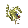
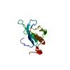
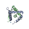
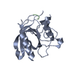
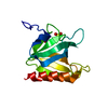
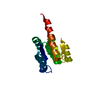
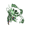
| Title | Crystal structure of the human Fe65-PTB1 domain | ||||||
 Components Components | Amyloid beta A4 precursor protein-binding family B member 1 | ||||||
 Keywords Keywords | PROTEIN BINDING / alpha-beta structure / phosphotyrosine binding domain | ||||||
| Function / homology |  Function and homology information Function and homology informationnegative regulation of cell cycle G1/S phase transition / proline-rich region binding / low-density lipoprotein particle receptor binding / smooth muscle contraction / axonogenesis / positive regulation of protein secretion / positive regulation of neuron projection development / lamellipodium / amyloid-beta binding / Recruitment and ATM-mediated phosphorylation of repair and signaling proteins at DNA double strand breaks ...negative regulation of cell cycle G1/S phase transition / proline-rich region binding / low-density lipoprotein particle receptor binding / smooth muscle contraction / axonogenesis / positive regulation of protein secretion / positive regulation of neuron projection development / lamellipodium / amyloid-beta binding / Recruitment and ATM-mediated phosphorylation of repair and signaling proteins at DNA double strand breaks / chromatin organization / growth cone / histone binding / molecular adaptor activity / transcription coactivator activity / nuclear speck / positive regulation of apoptotic process / apoptotic process / chromatin binding / DNA damage response / ubiquitin protein ligase binding / regulation of DNA-templated transcription / synapse / positive regulation of DNA-templated transcription / negative regulation of transcription by RNA polymerase II / signal transduction / endoplasmic reticulum / positive regulation of transcription by RNA polymerase II / nucleoplasm / nucleus / plasma membrane / cytoplasm Similarity search - Function | ||||||
| Biological species |  Homo sapiens (human) Homo sapiens (human) | ||||||
| Method |  X-RAY DIFFRACTION / SYNCHROTRON / SAD / Resolution: 2.2 Å X-RAY DIFFRACTION / SYNCHROTRON / SAD / Resolution: 2.2 Å | ||||||
 Authors Authors | Radzimanowski, J. / Ravaud, S. / Sinning, I. / Wild, K. | ||||||
 Citation Citation | Journal: J.Biol.Chem. / Year: 2008 Title: Crystal structure of the human Fe65-PTB1 domain. Authors: Radzimanowski, J. / Ravaud, S. / Schlesinger, S. / Koch, J. / Beyreuther, K. / Sinning, I. / Wild, K. #1: Journal: Acta Crystallogr.,Sect.F / Year: 2008 Title: Mercury-induced crystallization and SAD phasing of the human Fe65-PTB1 domain. Authors: Radzimanowski, J. / Ravaud, S. / Beyreuther, K. / Sinning, I. / Wild, K. | ||||||
| History |
|
- Structure visualization
| Structure viewer | Molecule: MolmilJmol/JSmol |
|---|
- Downloads & links
-Download
| PDBx/mmCIF format | 3d8d.cif.gz | 70.2 KB | Display | PDBx/mmCIF format |
|---|---|---|---|---|
| PDB format | pdb3d8d.ent.gz | 52 KB | Display | PDB format |
| PDBx/mmJSON format | 3d8d.json.gz | Tree view | PDBx/mmJSON format | |
| Others |  Other downloads Other downloads |
-Validation report
| Arichive directory | https://data.pdbj.org/pub/pdb/validation_reports/d8/3d8dftp://data.pdbj.org/pub/pdb/validation_reports/d8/3d8d | HTTPS FTP |
|---|
-Related structure data

-Links
 PDBj
PDBj


- Assembly
| Deposited unit | 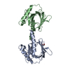
| ||||||||
|---|---|---|---|---|---|---|---|---|---|
| 1 | 
| ||||||||
| 2 | 
| ||||||||
| 3 | 
| ||||||||
| 4 | 
| ||||||||
| 5 | 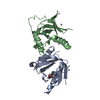
| ||||||||
| Unit cell |
|
-Components
| #1: Protein | Mass: 16782.211 Da / Num. of mol.: 2 / Fragment: Fe65-PTB1 domain Source method: isolated from a genetically manipulated source Source: (gene. exp.) Homo sapiens (human) / Gene: APBB1, FE65, RIR / Plasmid: pET24d / Production host:  #2: Chemical | ChemComp-HG /   Mass: 200.590 Da / Num. of mol.: 6 / Source method: obtained synthetically / Formula: Hg Mass: 200.590 Da / Num. of mol.: 6 / Source method: obtained synthetically / Formula: Hg#3: Chemical | ChemComp-EDO / |   Mass: 62.068 Da / Num. of mol.: 1 / Source method: obtained synthetically / Formula: C2H6O2 Mass: 62.068 Da / Num. of mol.: 1 / Source method: obtained synthetically / Formula: C2H6O2#4: Water | ChemComp-HOH / |  Mass: 18.015 Da / Num. of mol.: 130 / Source method: isolated from a natural source / Formula: H2O Mass: 18.015 Da / Num. of mol.: 130 / Source method: isolated from a natural source / Formula: H2O |
|---|
-Experimental details
-Experiment
| Experiment | Method: X-RAY DIFFRACTION / Number of used crystals: 1 |
|---|
- Sample preparation
| Crystal | Density Matthews: 2.18 Å3/Da / Density % sol: 43.52 % |
|---|---|
| Crystal grow | Temperature: 291 K / Method: vapor diffusion / pH: 7.5 Details: 100mM HEPES, 5% (v/v) ethylene glycol, 8% (w/v) PEG 3350, 1mM CH3HgCl, pH 7.5, VAPOR DIFFUSION, temperature 291K |
-Data collection
| Diffraction | Mean temperature: 100 K |
|---|---|
| Diffraction source | Source: SYNCHROTRON / Site: ESRF  / Beamline: ID23-1 / Wavelength: 1.0073 Å / Beamline: ID23-1 / Wavelength: 1.0073 Å |
| Detector | Type: ADSC QUANTUM 315 / Detector: CCD / Date: May 18, 2007 |
| Radiation | Protocol: SINGLE WAVELENGTH / Monochromatic (M) / Laue (L): M / Scattering type: x-ray |
| Radiation wavelength | Wavelength: 1.0073 Å / Relative weight: 1 |
| Reflection | Resolution: 2.2→83.9 Å / Num. obs: 15508 / % possible obs: 100 % / Observed criterion σ(F): 0 / Observed criterion σ(I): 0 / Redundancy: 6.2 % / Rmerge(I) obs: 0.067 / Net I/σ(I): 19.2 |
| Reflection shell | Resolution: 2.2→2.32 Å / Redundancy: 6.4 % / Rmerge(I) obs: 0.347 / Mean I/σ(I) obs: 4.6 / % possible all: 100 |
- Processing
| Software |
| ||||||||||||||||||||||||||||||||||||||||||||||||||||||||||||||||||||||||||||||||||||||||||
|---|---|---|---|---|---|---|---|---|---|---|---|---|---|---|---|---|---|---|---|---|---|---|---|---|---|---|---|---|---|---|---|---|---|---|---|---|---|---|---|---|---|---|---|---|---|---|---|---|---|---|---|---|---|---|---|---|---|---|---|---|---|---|---|---|---|---|---|---|---|---|---|---|---|---|---|---|---|---|---|---|---|---|---|---|---|---|---|---|---|---|---|
| Refinement | Method to determine structure: SAD / Resolution: 2.2→42 Å / Cor.coef. Fo:Fc: 0.944 / Cor.coef. Fo:Fc free: 0.92 / SU B: 5.935 / SU ML: 0.154 / Cross valid method: THROUGHOUT / σ(F): 0 / ESU R: 0.296 / ESU R Free: 0.225 / Stereochemistry target values: MAXIMUM LIKELIHOOD
| ||||||||||||||||||||||||||||||||||||||||||||||||||||||||||||||||||||||||||||||||||||||||||
| Solvent computation | Ion probe radii: 0.8 Å / Shrinkage radii: 0.8 Å / VDW probe radii: 1.2 Å / Solvent model: MASK | ||||||||||||||||||||||||||||||||||||||||||||||||||||||||||||||||||||||||||||||||||||||||||
| Displacement parameters | Biso mean: 35.469 Å2
| ||||||||||||||||||||||||||||||||||||||||||||||||||||||||||||||||||||||||||||||||||||||||||
| Refine analyze | Luzzati sigma a obs: 0.267 Å | ||||||||||||||||||||||||||||||||||||||||||||||||||||||||||||||||||||||||||||||||||||||||||
| Refinement step | Cycle: LAST / Resolution: 2.2→42 Å
| ||||||||||||||||||||||||||||||||||||||||||||||||||||||||||||||||||||||||||||||||||||||||||
| Refine LS restraints |
| ||||||||||||||||||||||||||||||||||||||||||||||||||||||||||||||||||||||||||||||||||||||||||
| LS refinement shell | Resolution: 2.2→2.257 Å / Total num. of bins used: 20
|
