[English] 日本語
 Yorodumi
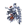
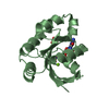
- PDB-1fho: Solution Structure of the PH Domain from the C. Elegans Muscle Pr... -
+ Open data
- Basic information
| Entry | Database: PDB / ID: 1fho | ||||||
|---|---|---|---|---|---|---|---|
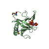
| Title | Solution Structure of the PH Domain from the C. Elegans Muscle Protein UNC-89 | ||||||
 Components Components | UNC-89 | ||||||
 Keywords Keywords | SIGNALING PROTEIN / Pleckstrin Homology domain / electrostatics / muscle / signal transduction | ||||||
| Function / homology |  Function and homology information Function and homology informationnematode pharyngeal gland morphogenesis / Synaptic adhesion-like molecules / regulation of skeletal muscle contraction by calcium ion signaling / MATH domain binding / striated muscle myosin thick filament assembly / positive regulation of sarcomere organization / positive regulation of locomotion / myosin filament assembly / positive regulation of protein localization to endoplasmic reticulum / positive regulation of striated muscle contraction ...nematode pharyngeal gland morphogenesis / Synaptic adhesion-like molecules / regulation of skeletal muscle contraction by calcium ion signaling / MATH domain binding / striated muscle myosin thick filament assembly / positive regulation of sarcomere organization / positive regulation of locomotion / myosin filament assembly / positive regulation of protein localization to endoplasmic reticulum / positive regulation of striated muscle contraction / skeletal muscle myosin thick filament assembly / A band / M band / sarcomere organization / phosphatase binding / guanyl-nucleotide exchange factor activity / small GTPase binding / intracellular protein localization / protein kinase activity / positive regulation of gene expression / ATP binding Similarity search - Function | ||||||
| Biological species |  | ||||||
| Method | SOLUTION NMR / Automated NOE assignment,Torsion angle dynamics, Cartesian simulated annealing. | ||||||
 Authors Authors | Blomberg, N. / Baraldi, E. / Sattler, M. / Saraste, M. / Nilges, M. | ||||||
 Citation Citation |  Journal: Structure Fold.Des. / Year: 2000 Journal: Structure Fold.Des. / Year: 2000Title: Structure of a PH domain from the C. elegans muscle protein UNC-89 suggests a novel function. Authors: Blomberg, N. / Baraldi, E. / Sattler, M. / Saraste, M. / Nilges, M. #1:  Journal: J.Biomol.NMR / Year: 1999 Journal: J.Biomol.NMR / Year: 1999Title: 1H, 15N, and 13C Resonance Assignment of the PH Domain from C. elegans UNC-89 Authors: Blomberg, N. / Sattler, M. / Nilges, M. #2:  Journal: Proteins / Year: 1999 Journal: Proteins / Year: 1999Title: Classification of Protein Sequences by Homology Modelling and Quantitative Analysis of Electrostatic Similarity Authors: Blomberg, N. / Gabdoulline, R.R. / Nilges, M. / Wade, R.C. #3:  Journal: Fold.Des. / Year: 1997 Journal: Fold.Des. / Year: 1997Title: Functional Diversity of PH Domains: an Exhaustive Modelling Study Authors: Blomberg, N. / Nilges, M. | ||||||
| History |
|
- Structure visualization
| Structure viewer | Molecule:  Molmil Molmil Jmol/JSmol Jmol/JSmol |
|---|
- Downloads & links
- Download
| PDBx/mmCIF format |  1fho.cif.gz 1fho.cif.gz | 1022.7 KB | Display |  PDBx/mmCIF format PDBx/mmCIF format |
|---|---|---|---|---|
| PDB format |  pdb1fho.ent.gz pdb1fho.ent.gz | 858.2 KB | Display |  PDB format PDB format |
| PDBx/mmJSON format |  1fho.json.gz 1fho.json.gz | Tree view |  PDBx/mmJSON format PDBx/mmJSON format | |
| Others |  Other downloads Other downloads |
-Validation report
| Summary document |  1fho_validation.pdf.gz 1fho_validation.pdf.gz | 356.5 KB | Display |  wwPDB validaton report wwPDB validaton report |
|---|---|---|---|---|
| Full document |  1fho_full_validation.pdf.gz 1fho_full_validation.pdf.gz | 581.8 KB | Display | |
| Data in XML |  1fho_validation.xml.gz 1fho_validation.xml.gz | 60.6 KB | Display | |
| Data in CIF |  1fho_validation.cif.gz 1fho_validation.cif.gz | 92.4 KB | Display | |
| Arichive directory |  https://data.pdbj.org/pub/pdb/validation_reports/fh/1fho https://data.pdbj.org/pub/pdb/validation_reports/fh/1fho ftp://data.pdbj.org/pub/pdb/validation_reports/fh/1fho ftp://data.pdbj.org/pub/pdb/validation_reports/fh/1fho | HTTPS FTP |
-Related structure data
| Similar structure data |
|---|
- Links
Links
- Assembly
| Deposited unit | 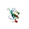
| |||||||||
|---|---|---|---|---|---|---|---|---|---|---|
| 1 |
| |||||||||
| NMR ensembles |
|
- Components
| #1: Protein | Mass: 14103.741 Da / Num. of mol.: 1 / Fragment: PLECKSTRIN HOMOLOGY (PH) DOMAIN Source method: isolated from a genetically manipulated source Source: (gene. exp.)   |
|---|
-Experimental details
-Experiment
| Experiment | Method: SOLUTION NMR | ||||||||||||||||||||
|---|---|---|---|---|---|---|---|---|---|---|---|---|---|---|---|---|---|---|---|---|---|
| NMR experiment |
| ||||||||||||||||||||
| NMR details | Text: Structure determined using triple resonance NMR techniques. |
- Sample preparation
| Details |
| ||||||||||||||||||||
|---|---|---|---|---|---|---|---|---|---|---|---|---|---|---|---|---|---|---|---|---|---|
| Sample conditions |
| ||||||||||||||||||||
| Crystal grow | *PLUS Method: other / Details: NMR |
-NMR measurement
| NMR spectrometer |
|
|---|
- Processing
| NMR software |
| ||||||||||||||||||||||||||||
|---|---|---|---|---|---|---|---|---|---|---|---|---|---|---|---|---|---|---|---|---|---|---|---|---|---|---|---|---|---|
| Refinement | Method: Automated NOE assignment,Torsion angle dynamics, Cartesian simulated annealing. Software ordinal: 1 Details: The structure was calculated automated NOE assignment and structure calculation using ARIA/CNS. Manual NOE assignments were added between cycles of automated assignment. The final structures ...Details: The structure was calculated automated NOE assignment and structure calculation using ARIA/CNS. Manual NOE assignments were added between cycles of automated assignment. The final structures were refined in a shell of explicit solvent. Data consisted of: 1230 unique NOE restaints; 44 phi restraints (3JHNHA scalar couplings, direct refinement against couplings); 41 1JHN residual dipolar couplings. Final ensemble was refined in explicit water. | ||||||||||||||||||||||||||||
| NMR representative | Selection criteria: lowest energy | ||||||||||||||||||||||||||||
| NMR ensemble | Conformer selection criteria: structures with the lowest energy Conformers calculated total number: 50 / Conformers submitted total number: 25 |
 Movie
Movie Controller




 PDBj
PDBj


 HSQC
HSQC