 Movie
Movie Controller


+ Open data

- Basic information
| Entry | Database: EMDB / ID: EMD-6211 | |||||||||
|---|---|---|---|---|---|---|---|---|---|---|
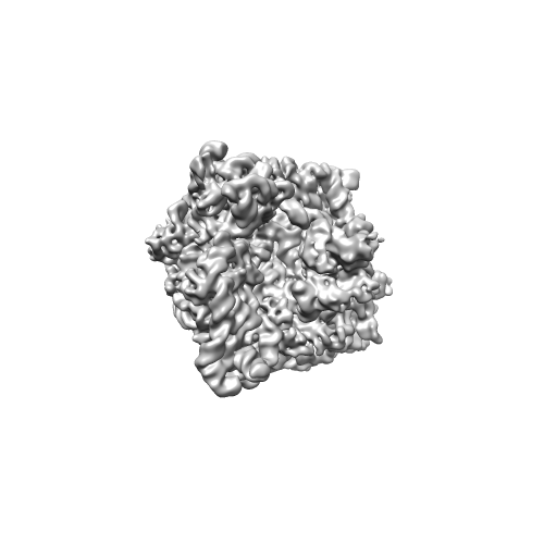
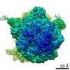
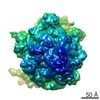
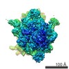
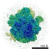
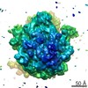
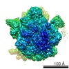
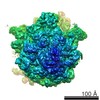
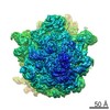
| Title | Cryo-EM structure of ribosomal protein S1 on the ribosome | |||||||||
 Map data Map data | Reconstruction of ErmCL-stalled E. coli ribosome containing density for the first domains of ribosomal protein S1 | |||||||||
 Sample Sample |
| |||||||||
 Keywords Keywords | Ribosome / Ribosomal protein S1 / Cryo-EM | |||||||||
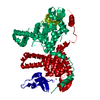
| Biological species |  | |||||||||
| Method | single particle reconstruction / cryo EM / Resolution: 9.5 Å | |||||||||
 Authors Authors | Byrgazov K / Grishkovskaya I / Arenz S / Coudevylle N / Temmel H / Wilson DN / Djinovic-Carugo K / Moll I | |||||||||
 Citation Citation | Journal: Nucleic Acids Res / Year: 2015 Title: Structural basis for the interaction of protein S1 with the Escherichia coli ribosome. Authors: Konstantin Byrgazov / Irina Grishkovskaya / Stefan Arenz / Nicolas Coudevylle / Hannes Temmel / Daniel N Wilson / Kristina Djinovic-Carugo / Isabella Moll /    Abstract: In Gram-negative bacteria, the multi-domain protein S1 is essential for translation initiation, as it recruits the mRNA and facilitates its localization in the decoding centre. In sharp contrast to ...In Gram-negative bacteria, the multi-domain protein S1 is essential for translation initiation, as it recruits the mRNA and facilitates its localization in the decoding centre. In sharp contrast to its functional importance, S1 is still lacking from the high-resolution structures available for Escherichia coli and Thermus thermophilus ribosomes and thus the molecular mechanism governing the S1-ribosome interaction has still remained elusive. Here, we present the structure of the N-terminal S1 domain D1 when bound to the ribosome at atomic resolution by using a combination of NMR, X-ray crystallography and cryo-electron microscopy. Together with biochemical assays, the structure reveals that S1 is anchored to the ribosome primarily via a stabilizing π-stacking interaction within the short but conserved N-terminal segment that is flexibly connected to domain D1. This interaction is further stabilized by salt bridges involving the zinc binding pocket of protein S2. Overall, this work provides one hitherto enigmatic piece in the 'ribosome puzzle', namely the detailed molecular insight into the topology of the S1-ribosome interface. Moreover, our data suggest novel mechanisms that have the potential to modulate protein synthesis in response to environmental cues by changing the affinity of S1 for the ribosome. | |||||||||
| History |
|
- Structure visualization
| Movie |
 Movie viewer Movie viewer |
|---|---|
| Structure viewer | EM map: SurfViewMolmilJmol/JSmol |
| Supplemental images |
- Downloads & links
-EMDB archive
| Map data | emd_6211.map.gz | 177.1 MB | EMDB map data format | |
|---|---|---|---|---|
| Header (meta data) | emd-6211-v30.xmlemd-6211.xml | 8.7 KB 8.7 KB | Display Display | EMDB header |
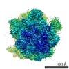
| Images |  emd_6211.png emd_6211.png | 61.5 KB | ||
| Archive directory |  http://ftp.pdbj.org/pub/emdb/structures/EMD-6211ftp://ftp.pdbj.org/pub/emdb/structures/EMD-6211 http://ftp.pdbj.org/pub/emdb/structures/EMD-6211ftp://ftp.pdbj.org/pub/emdb/structures/EMD-6211 | HTTPS FTP |
-Related structure data
-Links
| EMDB pages | EMDB (EBI/PDBe) / EMDataResource |
|---|---|
| Related items in Molecule of the Month |

-Map
| File | Download / File: emd_6211.map.gz / Format: CCP4 / Size: 185.7 MB / Type: IMAGE STORED AS FLOATING POINT NUMBER (4 BYTES) | ||||||||||||||||||||||||||||||||||||||||||||||||||||||||||||||||||||
|---|---|---|---|---|---|---|---|---|---|---|---|---|---|---|---|---|---|---|---|---|---|---|---|---|---|---|---|---|---|---|---|---|---|---|---|---|---|---|---|---|---|---|---|---|---|---|---|---|---|---|---|---|---|---|---|---|---|---|---|---|---|---|---|---|---|---|---|---|---|
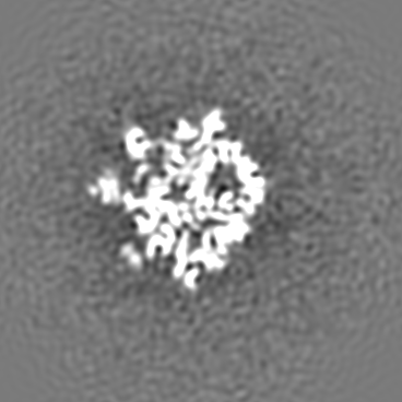
| Annotation | Reconstruction of ErmCL-stalled E. coli ribosome containing density for the first domains of ribosomal protein S1 | ||||||||||||||||||||||||||||||||||||||||||||||||||||||||||||||||||||
| Projections & slices | Image control
Images are generated by Spider. | ||||||||||||||||||||||||||||||||||||||||||||||||||||||||||||||||||||
| Voxel size | X=Y=Z: 1.0489 Å | ||||||||||||||||||||||||||||||||||||||||||||||||||||||||||||||||||||
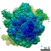
| Density |
| ||||||||||||||||||||||||||||||||||||||||||||||||||||||||||||||||||||
| Symmetry | Space group: 1 | ||||||||||||||||||||||||||||||||||||||||||||||||||||||||||||||||||||
| Details | EMDB XML:
CCP4 map header:
| ||||||||||||||||||||||||||||||||||||||||||||||||||||||||||||||||||||
 Z (Sec.)
 Y (Row.)
 X (Col.)


-Supplemental data
- Sample components
-Entire : Reconstruction of ErmCL-stalled E. coli ribosome containing densi...
| Entire | Name: Reconstruction of ErmCL-stalled E. coli ribosome containing density for the first domains of ribosomal protein S1 |
|---|---|
| Components |
|
-Supramolecule #1000: Reconstruction of ErmCL-stalled E. coli ribosome containing densi...
| Supramolecule | Name: Reconstruction of ErmCL-stalled E. coli ribosome containing density for the first domains of ribosomal protein S1 type: sample / ID: 1000 / Number unique components: 1 |
|---|
-Supramolecule #1: ErmCL-stalled ribosome
| Supramolecule | Name: ErmCL-stalled ribosome / type: complex / ID: 1 / Name.synonym: 70S ribosome / Recombinant expression: No / Database: NCBI / Ribosome-details: ribosome-prokaryote: ALL |
|---|---|
| Source (natural) | Organism: |
-Experimental details
-Structure determination
| Method | cryo EM |
|---|---|
 Processing Processing | single particle reconstruction |
| Aggregation state | particle |
-Sample preparation
| Buffer | pH: 7.4 / Details: 50 mM HEPES, 100 mM KOAc, 20 mM MgOAc |
|---|---|
| Grid | Details: 2 nm pre-coated Quantifoil R3/3 holey carbon supported grids |
| Vitrification | Cryogen name: ETHANE / Instrument: FEI VITROBOT MARK IV |
- Electron microscopy
| Microscope | FEI TITAN KRIOS |
|---|---|
| Date | May 21, 2012 |
| Image recording | Category: CCD / Film or detector model: TVIPS TEMCAM-F416 (4k x 4k) / Number real images: 6902 / Average electron dose: 20 e/Å2 |
| Electron beam | Acceleration voltage: 200 kV / Electron source:  FIELD EMISSION GUN FIELD EMISSION GUN |
| Electron optics | Illumination mode: SPOT SCAN / Imaging mode: BRIGHT FIELD / Cs: 2.7 mm / Nominal defocus max: 4.5 µm / Nominal defocus min: 1.2 µm / Nominal magnification: 75000 |
| Sample stage | Specimen holder model: FEI TITAN KRIOS AUTOGRID HOLDER |
| Experimental equipment |  Model: Titan Krios / Image courtesy: FEI Company |
-Image processing
| Details | CTF-parameter estimation in SPIDER TF ED, particle picking in Signature, image processing in SPIDER |
|---|---|
| CTF correction | Details: Defocus groups |
| Final reconstruction | Resolution.type: BY AUTHOR / Resolution: 9.5 Å / Resolution method: OTHER / Software - Name: SPIDER / Details: Final map was filtered to 9.5 Angstrom resolution. / Number images used: 128846 |
