ムービー
ムービー コントローラー


+ データを開く

- 基本情報
基本情報
| 登録情報 | データベース: EMDB / ID: EMD-2640 | |||||||||
|---|---|---|---|---|---|---|---|---|---|---|
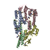
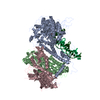
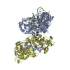
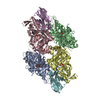
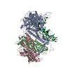
| タイトル | Negative-stain electron microscopy of the human GINS complex | |||||||||
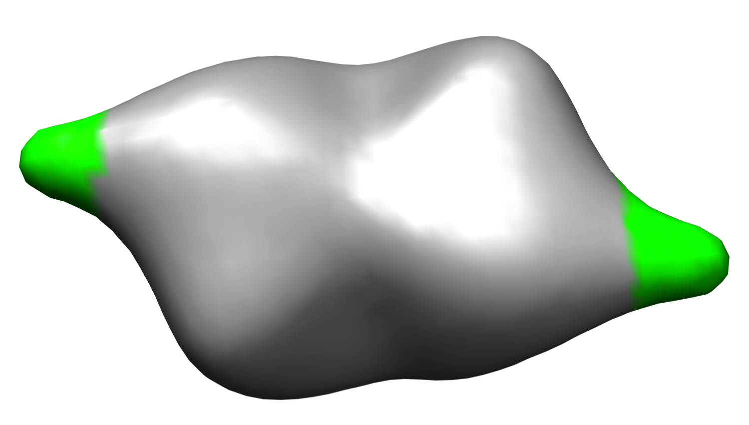
 マップデータ マップデータ | Reconstruction of the human GINS complex | |||||||||
 試料 試料 |
| |||||||||
 キーワード キーワード | DNA replication / CMG / GINS complex / Psf1 B domain | |||||||||
| 生物種 |  Homo sapiens (ヒト) Homo sapiens (ヒト) | |||||||||
| 手法 | 単粒子再構成法 / ネガティブ染色法 / 解像度: 30.0 Å | |||||||||
 データ登録者 データ登録者 | Carroni M / De March M / Krastanova I / Medagli B / Taylor IK / Brick P / Amenitsch H / Patwardhan A / Onesti S | |||||||||
 引用 引用 | ジャーナル: Sci Rep / 年: 2017 タイトル: New insights into the GINS complex explain the controversy between existing structural models. 著者: Marta Carroni / Matteo De March / Barbara Medagli / Ivet Krastanova / Ian A Taylor / Heinz Amenitsch / Hiroyuchi Araki / Francesca M Pisani / Ardan Patwardhan / Silvia Onesti /     要旨: GINS is a key component of eukaryotic replicative forks and is composed of four subunits (Sld5, Psf1, Psf2, Psf3). To explain the discrepancy between structural data from crystallography and electron ...GINS is a key component of eukaryotic replicative forks and is composed of four subunits (Sld5, Psf1, Psf2, Psf3). To explain the discrepancy between structural data from crystallography and electron microscopy (EM), we show that GINS is a compact tetramer in solution as observed in crystal structures, but also forms a double-tetrameric population, detectable by EM. This may represent an intermediate step towards the assembly of two replicative helicase complexes at origins, moving in opposite directions within the replication bubble. Reconstruction of the double-tetrameric form, combined with small-angle X-ray scattering data, allows the localisation of the B domain of the Psf1 subunit in the free GINS complex, which was not visible in previous studies and is essential for the formation of a functional replication fork. | |||||||||
| 履歴 |
|
- 構造の表示
構造の表示
| ムービー |
 ムービービューア ムービービューア |
|---|---|
| 構造ビューア | EMマップ: SurfViewMolmilJmol/JSmol |
| 添付画像 |
- ダウンロードとリンク
-EMDBアーカイブ
| マップデータ | emd_2640.map.gz | 96.6 KB | EMDBマップデータ形式 | |
|---|---|---|---|---|
| ヘッダ (付随情報) | emd-2640-v30.xmlemd-2640.xml | 10.4 KB 10.4 KB | 表示 表示 | EMDBヘッダ |
| 画像 |  emd_2640.png emd_2640.png | 342.3 KB | ||
| アーカイブディレクトリ |  http://ftp.pdbj.org/pub/emdb/structures/EMD-2640ftp://ftp.pdbj.org/pub/emdb/structures/EMD-2640 http://ftp.pdbj.org/pub/emdb/structures/EMD-2640ftp://ftp.pdbj.org/pub/emdb/structures/EMD-2640 | HTTPS FTP |
-検証レポート
| 文書・要旨 | emd_2640_validation.pdf.gz | 186.7 KB | 表示 | EMDB検証レポート |
|---|---|---|---|---|
| 文書・詳細版 | emd_2640_full_validation.pdf.gz | 185.9 KB | 表示 | |
| XML形式データ | emd_2640_validation.xml.gz | 4.4 KB | 表示 | |
| アーカイブディレクトリ | https://ftp.pdbj.org/pub/emdb/validation_reports/EMD-2640ftp://ftp.pdbj.org/pub/emdb/validation_reports/EMD-2640 | HTTPS FTP |
-関連構造データ


-リンク
| EMDBのページ | EMDB (EBI/PDBe) / EMDataResource |
|---|
-マップ
| ファイル | ダウンロード / ファイル: emd_2640.map.gz / 形式: CCP4 / 大きさ: 1.9 MB / タイプ: IMAGE STORED AS FLOATING POINT NUMBER (4 BYTES) | ||||||||||||||||||||||||||||||||||||||||||||||||||||||||||||
|---|---|---|---|---|---|---|---|---|---|---|---|---|---|---|---|---|---|---|---|---|---|---|---|---|---|---|---|---|---|---|---|---|---|---|---|---|---|---|---|---|---|---|---|---|---|---|---|---|---|---|---|---|---|---|---|---|---|---|---|---|---|
| 注釈 | Reconstruction of the human GINS complex | ||||||||||||||||||||||||||||||||||||||||||||||||||||||||||||
| 投影像・断面図 | 画像のコントロール
画像は Spider により作成 | ||||||||||||||||||||||||||||||||||||||||||||||||||||||||||||
| ボクセルのサイズ | X=Y=Z: 4 Å | ||||||||||||||||||||||||||||||||||||||||||||||||||||||||||||
| 密度 |
| ||||||||||||||||||||||||||||||||||||||||||||||||||||||||||||
| 対称性 | 空間群: 1 | ||||||||||||||||||||||||||||||||||||||||||||||||||||||||||||
| 詳細 | EMDB XML:
CCP4マップ ヘッダ情報:
| ||||||||||||||||||||||||||||||||||||||||||||||||||||||||||||
 Z (Sec.)
 Y (Row.)
 X (Col.)


-添付データ
- 試料の構成要素
-全体 : Human GINS complex
| 全体 | 名称: Human GINS complex |
|---|---|
| 要素 |
|
-超分子 #1000: Human GINS complex
| 超分子 | 名称: Human GINS complex / タイプ: sample / ID: 1000 詳細: The sample was in equilibrium with a monomeric form (single GINS tetramer) 集合状態: dimer of human GINS tetramers / Number unique components: 1 |
|---|---|
| 分子量 | 実験値: 200 KDa / 理論値: 200 KDa 手法: SEC-MALLS (size-exclusion chromatography coupled with multi angle laser light scattering) |
-分子 #1: GINS
| 分子 | 名称: GINS / タイプ: protein_or_peptide / ID: 1 / コピー数: 2 / 集合状態: dimer / 組換発現: Yes |
|---|---|
| 由来(天然) | 生物種: Homo sapiens (ヒト) / 別称: Human / 組織: all / 細胞: all / Organelle: nucleus / 細胞中の位置: nucleus |
| 分子量 | 実験値: 100 KDa / 理論値: 100 KDa |
| 組換発現 | 生物種:  |
-実験情報
-構造解析
| 手法 | ネガティブ染色法 |
|---|---|
 解析 解析 | 単粒子再構成法 |
| 試料の集合状態 | particle |
-試料調製
| 濃度 | 0.05 mg/mL |
|---|---|
| 緩衝液 | pH: 7.8 / 詳細: 20 mM TrisHCl pH 7.8, 150 mM NaCl, 0.5 mM TCEP |
| 染色 | タイプ: NEGATIVE 詳細: Protein adsorbed on carbon coated grids, stained with potassium phospho tungstate pH 7.0 for 4 minutes. |
| 凍結 | 凍結剤: NONE / 装置: OTHER |
- 電子顕微鏡法
| 顕微鏡 | FEI/PHILIPS CM200FEG |
|---|---|
| アライメント法 | Legacy - 非点収差: Objective lens astigmatism was corrected at 150,000 times magnification |
| 日付 | 2009年12月20日 |
| 撮影 | カテゴリ: CCD フィルム・検出器のモデル: TVIPS TEMCAM-F416 (4k x 4k) 平均電子線量: 20 e/Å2 |
| 電子線 | 加速電圧: 200 kV / 電子線源:  FIELD EMISSION GUN FIELD EMISSION GUN |
| 電子光学系 | 倍率(補正後): 50000 / 照射モード: FLOOD BEAM / 撮影モード: BRIGHT FIELD / Cs: 1.2 mm / 最大 デフォーカス(公称値): 3.0 µm / 最小 デフォーカス(公称値): 1.0 µm / 倍率(公称値): 50000 |
| 試料ステージ | 試料ホルダーモデル: SIDE ENTRY, EUCENTRIC |
-画像解析
| CTF補正 | 詳細: entire frame |
|---|---|
| 最終 再構成 | 想定した対称性 - 点群: C2 (2回回転対称) / アルゴリズム: OTHER / 解像度のタイプ: BY AUTHOR / 解像度: 30.0 Å / 解像度の算出法: OTHER / ソフトウェア - 名称: IMAGIC, Spider / 使用した粒子像数: 14390 |
-原子モデル構築 1
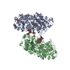
| 初期モデル | PDB ID: Chain - #0 - Chain ID: E / Chain - #1 - Chain ID: F / Chain - #2 - Chain ID: G / Chain - #3 - Chain ID: H / Chain - #4 - Chain ID: I / Chain - #5 - Chain ID: J / Chain - #6 - Chain ID: K / Chain - #7 - Chain ID: L |
|---|---|
| ソフトウェア | 名称: Chimera |
| 詳細 | A dimer of tetramers observed in the crystal was fitted. |
| 精密化 | 空間: REAL / プロトコル: RIGID BODY FIT |