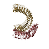
 Movie
Movie Controller


+ Open data

- Basic information
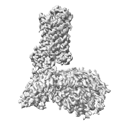
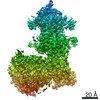
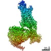
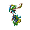
| Entry | Database: EMDB / ID: EMD-25819 | ||||||||||||
|---|---|---|---|---|---|---|---|---|---|---|---|---|---|
| Title | Lysophosphatidic acid receptor 1-Gi complex bound to LPA | ||||||||||||
 Map data Map data | |||||||||||||
 Sample Sample |
| ||||||||||||
 Keywords Keywords | GPCR / complex / lipid / MEMBRANE PROTEIN | ||||||||||||
| Function / homology |  Function and homology information Function and homology informationAdenylate cyclase inhibitory pathway / cellular response to 1-oleoyl-sn-glycerol 3-phosphate / lysophosphatidic acid receptor activity / positive regulation of smooth muscle cell chemotaxis / Adrenaline,noradrenaline inhibits insulin secretion / ADP signalling through P2Y purinoceptor 12 / adenylate cyclase regulator activity / lysophosphatidic acid binding / Lysosphingolipid and LPA receptors / Extra-nuclear estrogen signaling ...Adenylate cyclase inhibitory pathway / cellular response to 1-oleoyl-sn-glycerol 3-phosphate / lysophosphatidic acid receptor activity / positive regulation of smooth muscle cell chemotaxis / Adrenaline,noradrenaline inhibits insulin secretion / ADP signalling through P2Y purinoceptor 12 / adenylate cyclase regulator activity / lysophosphatidic acid binding / Lysosphingolipid and LPA receptors / Extra-nuclear estrogen signaling / Olfactory Signaling Pathway / Sensory perception of sweet, bitter, and umami (glutamate) taste / Synthesis, secretion, and inactivation of Glucagon-like Peptide-1 (GLP-1) / negative regulation of cilium assembly / corpus callosum development / G alpha (i) signalling events / Activation of the phototransduction cascade / oligodendrocyte development / cellular response to oxygen levels / optic nerve development / negative regulation of synaptic transmission / regulation of synaptic vesicle cycle / GTPase activating protein binding / Activation of G protein gated Potassium channels / G-protein activation / G beta:gamma signalling through PI3Kgamma / Prostacyclin signalling through prostacyclin receptor / G beta:gamma signalling through PLC beta / ADP signalling through P2Y purinoceptor 1 / Thromboxane signalling through TP receptor / Presynaptic function of Kainate receptors / G beta:gamma signalling through CDC42 / Inhibition of voltage gated Ca2+ channels via Gbeta/gamma subunits / G alpha (12/13) signalling events / Glucagon-type ligand receptors / G beta:gamma signalling through BTK / ADP signalling through P2Y purinoceptor 12 / Adrenaline,noradrenaline inhibits insulin secretion / Cooperation of PDCL (PhLP1) and TRiC/CCT in G-protein beta folding / Ca2+ pathway / G alpha (z) signalling events / Thrombin signalling through proteinase activated receptors (PARs) / Extra-nuclear estrogen signaling / G alpha (s) signalling events / G alpha (q) signalling events / regulation of postsynaptic neurotransmitter receptor internalization / positive regulation of dendritic spine development / Glucagon-like Peptide-1 (GLP1) regulates insulin secretion / G alpha (i) signalling events / neurotransmitter receptor localization to postsynaptic specialization membrane / High laminar flow shear stress activates signaling by PIEZO1 and PECAM1:CDH5:KDR in endothelial cells / Vasopressin regulates renal water homeostasis via Aquaporins / positive regulation of Rho protein signal transduction / G-protein alpha-subunit binding / negative regulation of cAMP/PKA signal transduction / centriolar satellite / positive regulation of stress fiber assembly / adenylate cyclase inhibitor activity / myelination / positive regulation of protein localization to cell cortex / T cell migration / neurogenesis / D2 dopamine receptor binding / adenylate cyclase-inhibiting serotonin receptor signaling pathway / cerebellum development / G protein-coupled serotonin receptor binding / cellular response to forskolin / regulation of mitotic spindle organization / dendritic shaft / chemokine-mediated signaling pathway / cell chemotaxis / PDZ domain binding / response to prostaglandin E / positive regulation of cholesterol biosynthetic process / G protein-coupled receptor binding / GABA-ergic synapse / G protein-coupled receptor activity / G-protein beta/gamma-subunit complex binding / adenylate cyclase-modulating G protein-coupled receptor signaling pathway / adenylate cyclase-inhibiting G protein-coupled receptor signaling pathway / photoreceptor disc membrane / GDP binding / regulation of cell shape / negative regulation of neuron projection development / cellular response to catecholamine stimulus / adenylate cyclase-activating dopamine receptor signaling pathway / cellular response to prostaglandin E stimulus / heterotrimeric G-protein complex / G-protein beta-subunit binding / positive regulation of cytosolic calcium ion concentration / sensory perception of taste / signaling receptor complex adaptor activity / adenylate cyclase-activating G protein-coupled receptor signaling pathway / retina development in camera-type eye / GTPase binding / presynaptic membrane / G protein activity / midbody / cell cortex / G alpha (i) signalling events Similarity search - Function | ||||||||||||
| Biological species |   Homo sapiens (human) / Homo sapiens (human) / | ||||||||||||
| Method | single particle reconstruction / cryo EM / Resolution: 2.83 Å | ||||||||||||
 Authors Authors | Liu S / Paknejad N | ||||||||||||
| Funding support |  United States, 3 items United States, 3 items
| ||||||||||||
 Citation Citation | Journal: Nat Commun / Year: 2022 Title: Differential activation mechanisms of lipid GPCRs by lysophosphatidic acid and sphingosine 1-phosphate. Authors: Shian Liu / Navid Paknejad / Lan Zhu / Yasuyuki Kihara / Manisha Ray / Jerold Chun / Wei Liu / Richard K Hite / Xin-Yun Huang / Abstract: Lysophospholipids are bioactive lipids and can signal through G-protein-coupled receptors (GPCRs). The best studied lysophospholipids are lysophosphatidic acid (LPA) and sphingosine 1-phosphate (S1P). ...Lysophospholipids are bioactive lipids and can signal through G-protein-coupled receptors (GPCRs). The best studied lysophospholipids are lysophosphatidic acid (LPA) and sphingosine 1-phosphate (S1P). The mechanisms of lysophospholipid recognition by an active GPCR, and the activations of lysophospholipid GPCR-G-protein complexes remain unclear. Here we report single-particle cryo-EM structures of human S1P receptor 1 (S1P) and heterotrimeric G complexes formed with bound S1P or the multiple sclerosis (MS) treatment drug Siponimod, as well as human LPA receptor 1 (LPA) and G complexes in the presence of LPA. Our structural and functional data provide insights into how LPA and S1P adopt different conformations to interact with their cognate GPCRs, the selectivity of the homologous lipid GPCRs for S1P versus LPA, and the different activation mechanisms of these GPCRs by LPA and S1P. Our studies also reveal specific optimization strategies to improve the MS-treating S1P-targeting drugs. | ||||||||||||
| History |
|
- Structure visualization
| Movie |
Movie viewer |
|---|---|
| Structure viewer | EM map: SurfViewMolmilJmol/JSmol |
| Supplemental images |
- Downloads & links
-EMDB archive
| Map data | emd_25819.map.gz | 10.1 MB | EMDB map data format | |
|---|---|---|---|---|
| Header (meta data) | emd-25819-v30.xmlemd-25819.xml | 14.1 KB 14.1 KB | Display Display | EMDB header |
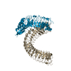
| Images |  emd_25819.png emd_25819.png | 89.4 KB | ||
| Filedesc metadata | emd-25819.cif.gz | 6.1 KB | ||
| Archive directory |  http://ftp.pdbj.org/pub/emdb/structures/EMD-25819ftp://ftp.pdbj.org/pub/emdb/structures/EMD-25819 http://ftp.pdbj.org/pub/emdb/structures/EMD-25819ftp://ftp.pdbj.org/pub/emdb/structures/EMD-25819 | HTTPS FTP |
-Related structure data
| Related structure data | 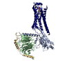 7td0MC 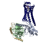 7td1C 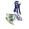 7td2C  7td3C  7td4C M: atomic model generated by this map C: citing same article ( |
|---|---|
| Similar structure data |

-Links
| EMDB pages | EMDB (EBI/PDBe) / EMDataResource |
|---|---|
| Related items in Molecule of the Month |


-Map
| File | Download / File: emd_25819.map.gz / Format: CCP4 / Size: 64 MB / Type: IMAGE STORED AS FLOATING POINT NUMBER (4 BYTES) | ||||||||||||||||||||||||||||||||||||||||||||||||||||||||||||||||||||
|---|---|---|---|---|---|---|---|---|---|---|---|---|---|---|---|---|---|---|---|---|---|---|---|---|---|---|---|---|---|---|---|---|---|---|---|---|---|---|---|---|---|---|---|---|---|---|---|---|---|---|---|---|---|---|---|---|---|---|---|---|---|---|---|---|---|---|---|---|---|
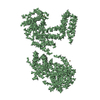
| Projections & slices | Image control
Images are generated by Spider. | ||||||||||||||||||||||||||||||||||||||||||||||||||||||||||||||||||||
| Voxel size | X=Y=Z: 1.064 Å | ||||||||||||||||||||||||||||||||||||||||||||||||||||||||||||||||||||
| Density |
| ||||||||||||||||||||||||||||||||||||||||||||||||||||||||||||||||||||
| Symmetry | Space group: 1 | ||||||||||||||||||||||||||||||||||||||||||||||||||||||||||||||||||||
| Details | EMDB XML:
CCP4 map header:
| ||||||||||||||||||||||||||||||||||||||||||||||||||||||||||||||||||||
 X (Sec.)
 Y (Row.)
 Z (Col.)


-Supplemental data
- Sample components
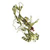
-Entire : complex of Lysophosphatidic Acid Receptor 1 with G-protein and LPA
| Entire | Name: complex of Lysophosphatidic Acid Receptor 1 with G-protein and LPA |
|---|---|
| Components |
|
-Supramolecule #1: complex of Lysophosphatidic Acid Receptor 1 with G-protein and LPA
| Supramolecule | Name: complex of Lysophosphatidic Acid Receptor 1 with G-protein and LPA type: complex / ID: 1 / Parent: 0 / Macromolecule list: #1-#4 |
|---|---|
| Source (natural) | Organism: |
-Macromolecule #1: Lysophosphatidic acid receptor 1
| Macromolecule | Name: Lysophosphatidic acid receptor 1 / type: protein_or_peptide / ID: 1 / Number of copies: 1 / Enantiomer: LEVO |
|---|---|
| Source (natural) | Organism: Homo sapiens (human) |
| Molecular weight | Theoretical: 39.734605 KDa |
| Recombinant expression | Organism:   Spodoptera frugiperda (fall armyworm) Spodoptera frugiperda (fall armyworm) |
| Sequence | String: DYKDDDDKAA AAAISTSIPV ISQPQFTAMN EPQCFYNESI AFFYNRSGKH LATEWNTVSK LVMGLGITVC IFIMLANLLV MVAIYVNRR FHFPIYYLMA NLAAADFFAG LAYFYLMFNT GPNTRRLTVS TWLLRQGLID TSLTASVANL LAIAIERHIT V FRMQLHTR ...String: DYKDDDDKAA AAAISTSIPV ISQPQFTAMN EPQCFYNESI AFFYNRSGKH LATEWNTVSK LVMGLGITVC IFIMLANLLV MVAIYVNRR FHFPIYYLMA NLAAADFFAG LAYFYLMFNT GPNTRRLTVS TWLLRQGLID TSLTASVANL LAIAIERHIT V FRMQLHTR MSNRRVVVVI VVIWTMAIVM GAIPSVGWNC ICDIENCSNM APLYSDSYLV FWAIFNLVTF VVMVVLYAHI FG YVRQRTM RMSRHSSGPR RNRDTMMSLL KTVVIVLGAF IICWTPGLVL LLLDVCCPQC DVLAYEKFFL LLAEFNSAMN PII YSYRDK EMSATFRQIL CCQRSENPTG PTEG UniProtKB: Lysophosphatidic acid receptor 1 |
-Macromolecule #2: Guanine nucleotide-binding protein G(I)/G(S)/G(T) subunit beta-1
| Macromolecule | Name: Guanine nucleotide-binding protein G(I)/G(S)/G(T) subunit beta-1 type: protein_or_peptide / ID: 2 / Number of copies: 1 / Enantiomer: LEVO |
|---|---|
| Source (natural) | Organism: |
| Molecular weight | Theoretical: 37.41693 KDa |
| Recombinant expression | Organism: Spodoptera frugiperda (fall armyworm) |
| Sequence | String: MSELDQLRQE AEQLKNQIRD ARKACADATL SQITNNIDPV GRIQMRTRRT LRGHLAKIYA MHWGTDSRLL VSASQDGKLI IWDSYTTNK VHAIPLRSSW VMTCAYAPSG NYVACGGLDN ICSIYNLKTR EGNVRVSREL AGHTGYLSCC RFLDDNQIVT S SGDTTCAL ...String: MSELDQLRQE AEQLKNQIRD ARKACADATL SQITNNIDPV GRIQMRTRRT LRGHLAKIYA MHWGTDSRLL VSASQDGKLI IWDSYTTNK VHAIPLRSSW VMTCAYAPSG NYVACGGLDN ICSIYNLKTR EGNVRVSREL AGHTGYLSCC RFLDDNQIVT S SGDTTCAL WDIETGQQTT TFTGHTGDVM SLSLAPDTRL FVSGACDASA KLWDVREGMC RQTFTGHESD INAICFFPNG NA FATGSDD ATCRLFDLRA DQELMTYSHD NIICGITSVS FSKSGRLLLA GYDDFNCNVW DALKADRAGV LAGHDNRVSC LGV TDDGMA VATGSWDSFL KIWN UniProtKB: Guanine nucleotide-binding protein G(I)/G(S)/G(T) subunit beta-1 |
-Macromolecule #3: Guanine nucleotide-binding protein G(i) subunit alpha-1
| Macromolecule | Name: Guanine nucleotide-binding protein G(i) subunit alpha-1 type: protein_or_peptide / ID: 3 / Number of copies: 1 / Enantiomer: LEVO |
|---|---|
| Source (natural) | Organism: |
| Molecular weight | Theoretical: 43.16307 KDa |
| Recombinant expression | Organism:  |
| Sequence | String: MGSSHHHHHH SSGLEVLFQG PHMASMGCTL SAEDKAAVER SKMIDRNLRE DGEKAAREVK LLLLGAGESG KSTIVKQMKI IHEAGYSEE ECKQYKAVVY SNTIQSIIAI IRAMGRLKID FGDSARADDA RQLFVLAGAA EEGFMTAELA GVIKRLWKDS G VQACFNRS ...String: MGSSHHHHHH SSGLEVLFQG PHMASMGCTL SAEDKAAVER SKMIDRNLRE DGEKAAREVK LLLLGAGESG KSTIVKQMKI IHEAGYSEE ECKQYKAVVY SNTIQSIIAI IRAMGRLKID FGDSARADDA RQLFVLAGAA EEGFMTAELA GVIKRLWKDS G VQACFNRS REYQLNDSAA YYLNDLDRIA QPNYIPTQQD VLRTRVKTTG IVETHFTFKD LHFKMFDVGA QRSERKKWIH CF EGVTAII FCVALSDYDL VLAEDEEMNR MHESMKLFDS ICNNKWFTDT SIILFLNKKD LFEEKIKKSP LTICYPEYAG SNT YEEAAA YIQCQFEDLN KRKDTKEIYT HFTCATDTKN VQFVFDAVTD VIIKNNLKDC GLF UniProtKB: Guanine nucleotide-binding protein G(i) subunit alpha-1 |
-Macromolecule #4: Guanine nucleotide-binding protein G(I)/G(S)/G(O) subunit gamma-2
| Macromolecule | Name: Guanine nucleotide-binding protein G(I)/G(S)/G(O) subunit gamma-2 type: protein_or_peptide / ID: 4 / Number of copies: 1 / Enantiomer: LEVO |
|---|---|
| Source (natural) | Organism: |
| Molecular weight | Theoretical: 7.845078 KDa |
| Recombinant expression | Organism: Spodoptera frugiperda (fall armyworm) |
| Sequence | String: MASNNTASIA QARKLVEQLK MEANIDRIKV SKAAADLMAY CEAHAKEDPL LTPVPASENP FREKKFFSAI L UniProtKB: Guanine nucleotide-binding protein G(I)/G(S)/G(O) subunit gamma-2 |
-Macromolecule #5: (2R)-2-hydroxy-3-(phosphonooxy)propyl (9E)-octadec-9-enoate
| Macromolecule | Name: (2R)-2-hydroxy-3-(phosphonooxy)propyl (9E)-octadec-9-enoate type: ligand / ID: 5 / Number of copies: 1 / Formula: NKP |
|---|---|
| Molecular weight | Theoretical: 436.52 Da |
| Chemical component information | 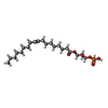 ChemComp-NKP: |
-Experimental details
-Structure determination
| Method | cryo EM |
|---|---|
 Processing Processing | single particle reconstruction |
| Aggregation state | particle |
-Sample preparation
| Buffer | pH: 7 |
|---|---|
| Vitrification | Cryogen name: ETHANE |
- Electron microscopy
| Microscope | FEI TITAN KRIOS |
|---|---|
| Image recording | Film or detector model: GATAN K3 (6k x 4k) / Average electron dose: 28.2 e/Å2 |
| Electron beam | Acceleration voltage: 300 kV / Electron source:  FIELD EMISSION GUN FIELD EMISSION GUN |
| Electron optics | Illumination mode: FLOOD BEAM / Imaging mode: BRIGHT FIELD / Nominal defocus max: 2.2 µm / Nominal defocus min: 0.8 µm |
| Experimental equipment |  Model: Titan Krios / Image courtesy: FEI Company |
-Image processing
| Startup model | Type of model: NONE |
|---|---|
| Final reconstruction | Resolution.type: BY AUTHOR / Resolution: 2.83 Å / Resolution method: FSC 0.143 CUT-OFF / Number images used: 1588791 |
| Initial angle assignment | Type: MAXIMUM LIKELIHOOD |
| Final angle assignment | Type: MAXIMUM LIKELIHOOD |
