 Movie
Movie Controller

[English] 日本語
 Yorodumi
- EMDB-2491: Recruitment Pathways in the Yeast Exosome by Electron Microscopy -
+ Open data

- Basic information
| Entry | Database: EMDB / ID: EMD-2491 | |||||||||
|---|---|---|---|---|---|---|---|---|---|---|
| Title | Recruitment Pathways in the Yeast Exosome by Electron Microscopy | |||||||||
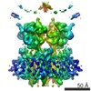
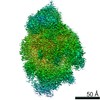
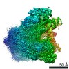
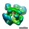
 Map data Map data | Exosome with short ssRNA | |||||||||
 Sample Sample |
| |||||||||
 Keywords Keywords | exosome / RNA degradation route | |||||||||
| Biological species |  | |||||||||
| Method | single particle reconstruction / negative staining / Resolution: 25.0 Å | |||||||||
 Authors Authors | Liu J-J / Bratkowski MA / Liu XQ / Niu C-Y / Ke AL / Wang H-W | |||||||||
 Citation Citation | Journal: Nat Struct Mol Biol / Year: 2014 Title: Visualization of distinct substrate-recruitment pathways in the yeast exosome by EM. Authors: Jun-Jie Liu / Matthew A Bratkowski / Xueqi Liu / Chu-Ya Niu / Ailong Ke / Hong-Wei Wang /   Abstract: The eukaryotic exosome is a multisubunit complex typically composed of a catalytically inactive core and the Rrp44 protein, which contains 3'-to-5' exo- and endo-RNase activities. RNA substrates have ...The eukaryotic exosome is a multisubunit complex typically composed of a catalytically inactive core and the Rrp44 protein, which contains 3'-to-5' exo- and endo-RNase activities. RNA substrates have been shown to be recruited through the core to reach Rrp44's exo-RNase (EXO) site. Using single-particle EM and biochemical analysis, we provide visual evidence that two distinct substrate-recruitment pathways exist. In the through-core route, channeling of the single-stranded substrates from the core to Rrp44 induces a characteristic conformational change in Rrp44. In the alternative direct-access route, this conformational change does not take place, and the RNA substrate is visualized to avoid the core and enter Rrp44's EXO site directly. Our results provide mechanistic explanations for several RNA processing scenarios by the eukaryotic exosome and indicate substrate-specific modes of degradation by this complex. | |||||||||
| History |
|
- Structure visualization
| Movie |
 Movie viewer Movie viewer |
|---|---|
| Structure viewer | EM map: SurfViewMolmilJmol/JSmol |
| Supplemental images |

- Downloads & links
-EMDB archive
| Map data | emd_2491.map.gz | 1.4 MB | EMDB map data format | |
|---|---|---|---|---|
| Header (meta data) | emd-2491-v30.xmlemd-2491.xml | 16.2 KB 16.2 KB | Display Display | EMDB header |
| Images | 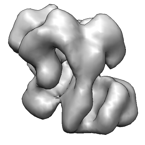 2491-RNA08-RE.png 2491-RNA08-RE.png | 64.3 KB | ||
| Archive directory |  http://ftp.pdbj.org/pub/emdb/structures/EMD-2491ftp://ftp.pdbj.org/pub/emdb/structures/EMD-2491 http://ftp.pdbj.org/pub/emdb/structures/EMD-2491ftp://ftp.pdbj.org/pub/emdb/structures/EMD-2491 | HTTPS FTP |
-Validation report
| Summary document | emd_2491_validation.pdf.gz | 219.6 KB | Display | EMDB validaton report |
|---|---|---|---|---|
| Full document | emd_2491_full_validation.pdf.gz | 218.7 KB | Display | |
| Data in XML | emd_2491_validation.xml.gz | 4.7 KB | Display | |
| Arichive directory | https://ftp.pdbj.org/pub/emdb/validation_reports/EMD-2491ftp://ftp.pdbj.org/pub/emdb/validation_reports/EMD-2491 | HTTPS FTP |
-Related structure data
| Related structure data |  2492C  2493C  2494C 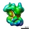 2495C 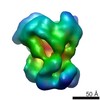 2496C 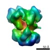 2497C 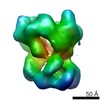 2498C 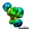 2499C 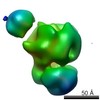 2500C 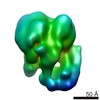 2501C 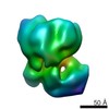 2502C 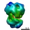 2522C C: citing same article ( |
|---|---|
| Similar structure data |
-Links
| EMDB pages | EMDB (EBI/PDBe) / EMDataResource |
|---|
-Map
| File | Download / File: emd_2491.map.gz / Format: CCP4 / Size: 2.7 MB / Type: IMAGE STORED AS FLOATING POINT NUMBER (4 BYTES) | ||||||||||||||||||||||||||||||||||||||||||||||||||||||||||||||||||||
|---|---|---|---|---|---|---|---|---|---|---|---|---|---|---|---|---|---|---|---|---|---|---|---|---|---|---|---|---|---|---|---|---|---|---|---|---|---|---|---|---|---|---|---|---|---|---|---|---|---|---|---|---|---|---|---|---|---|---|---|---|---|---|---|---|---|---|---|---|---|
| Annotation | Exosome with short ssRNA | ||||||||||||||||||||||||||||||||||||||||||||||||||||||||||||||||||||
| Projections & slices | Image control
Images are generated by Spider. | ||||||||||||||||||||||||||||||||||||||||||||||||||||||||||||||||||||
| Voxel size | X=Y=Z: 4.4 Å | ||||||||||||||||||||||||||||||||||||||||||||||||||||||||||||||||||||
| Density |
| ||||||||||||||||||||||||||||||||||||||||||||||||||||||||||||||||||||
| Symmetry | Space group: 1 | ||||||||||||||||||||||||||||||||||||||||||||||||||||||||||||||||||||
| Details | EMDB XML:
CCP4 map header:
| ||||||||||||||||||||||||||||||||||||||||||||||||||||||||||||||||||||
 Z (Sec.)
 Y (Row.)
 X (Col.)


-Supplemental data
- Sample components
+Entire : Rrp44-Exosome incubated with RNA08
+Supramolecule #1000: Rrp44-Exosome incubated with RNA08
+Macromolecule #1: Rrp44
+Macromolecule #2: Rrp43
+Macromolecule #3: Rrp4
+Macromolecule #4: Csl4
+Macromolecule #5: Rrp45
+Macromolecule #6: Rrp46-TAP
+Macromolecule #7: Rrp41
+Macromolecule #8: Rrp42
+Macromolecule #9: Mtr3
+Macromolecule #10: Rrp40
+Macromolecule #11: RNA08
-Experimental details
-Structure determination
| Method | negative staining |
|---|---|
 Processing Processing | single particle reconstruction |
| Aggregation state | particle |
-Sample preparation
| Concentration | 0.01 mg/mL |
|---|---|
| Buffer | pH: 8 / Details: 150mM NaCl, 50mM Tris-HCL,1mM DTT |
| Staining | Type: NEGATIVE Details: All samples were diluted at a final concentration of ~80 nM of the exosome in the non-digestive buffer and negatively stained in 2% (w/v) uranyl acetate solution following the standard deep ...Details: All samples were diluted at a final concentration of ~80 nM of the exosome in the non-digestive buffer and negatively stained in 2% (w/v) uranyl acetate solution following the standard deep stain procedure on holey-carbon coated EM copper grids covered with a thin layer of continuous carbon |
| Grid | Details: 300 mesh continuous carbon |
| Vitrification | Cryogen name: NONE / Instrument: OTHER |
- Electron microscopy
| Microscope | FEI TECNAI F20 |
|---|---|
| Specialist optics | Energy filter - Name: FEI |
| Date | Sep 10, 2012 |
| Image recording | Category: CCD / Film or detector model: GENERIC GATAN (4k x 4k) / Number real images: 300 / Average electron dose: 30 e/Å2 |
| Electron beam | Acceleration voltage: 200 kV / Electron source:  FIELD EMISSION GUN FIELD EMISSION GUN |
| Electron optics | Illumination mode: FLOOD BEAM / Imaging mode: BRIGHT FIELD / Cs: 2.7 mm |
| Sample stage | Specimen holder model: SIDE ENTRY, EUCENTRIC |
| Experimental equipment |  Model: Tecnai F20 / Image courtesy: FEI Company |
-Image processing
| Details | Briefly, we performed iterative multi-variant statistical analysis (MSA) and multi-reference alignment (MRA) cycles on the particle stacks using IMAGIC-4D to obtain 2D class averages as described previously36. We performed 3D reconstruction of all the samples using projection-matching refinement of particle stacks against the same initial model of apo-RE (EMD1439) low-pass filtered to 70 A in SPIDER39 as described previously36. The resolution of final reconstruction was estimated using Fourier Shell Correlation (FSC) algorithm at a criteria of 0.5 |
|---|---|
| Final reconstruction | Applied symmetry - Point group: C1 (asymmetric) / Resolution.type: BY AUTHOR / Resolution: 25.0 Å / Resolution method: FSC 0.5 CUT-OFF / Software - Name: SPIDER / Number images used: 30000 |
| Final angle assignment | Details: SPIDER: theta 90 degrees, phi 359.9 degrees |
| Final two d classification | Number classes: 600 |
