 ムービー
ムービー コントローラー


+ データを開く

- 基本情報
基本情報
| 登録情報 | データベース: EMDB / ID: EMD-2450 | |||||||||
|---|---|---|---|---|---|---|---|---|---|---|
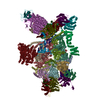
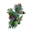
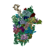
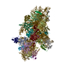
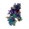
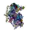
| タイトル | Cryo-EM map of the CSFV IRES in complex with the small ribosomal 40S subunit and DHX29 | |||||||||
 マップデータ マップデータ | Reconstruction of a mutant Classical Swine Fever Virus IRES bound to the Rabbit 40S subunit and DHX29. | |||||||||
 試料 試料 |
| |||||||||
 キーワード キーワード | Internal Ribosomal Entry Site / 5'-end independent initiation / HCV-like IRES | |||||||||
| 生物種 |   Classical swine fever virus (豚コレラウイルス) / Classical swine fever virus (豚コレラウイルス) /  Homo sapiens (ヒト) Homo sapiens (ヒト) | |||||||||
| 手法 | 単粒子再構成法 / クライオ電子顕微鏡法 / 解像度: 8.5 Å | |||||||||
 データ登録者 データ登録者 | Hashem Y / des Georges A / Dhote A / Langlois R / Liao HY / Grassucci RA / Pestova TV / Hellen CUT / Frank J | |||||||||
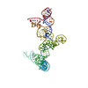
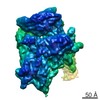
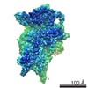
 引用 引用 | ジャーナル: Nature / 年: 2013 タイトル: Hepatitis-C-virus-like internal ribosome entry sites displace eIF3 to gain access to the 40S subunit. 著者: Yaser Hashem / Amedee des Georges / Vidya Dhote / Robert Langlois / Hstau Y Liao / Robert A Grassucci / Tatyana V Pestova / Christopher U T Hellen / Joachim Frank /  要旨: Hepatitis C virus (HCV) and classical swine fever virus (CSFV) messenger RNAs contain related (HCV-like) internal ribosome entry sites (IRESs) that promote 5'-end independent initiation of ...Hepatitis C virus (HCV) and classical swine fever virus (CSFV) messenger RNAs contain related (HCV-like) internal ribosome entry sites (IRESs) that promote 5'-end independent initiation of translation, requiring only a subset of the eukaryotic initiation factors (eIFs) needed for canonical initiation on cellular mRNAs. Initiation on HCV-like IRESs relies on their specific interaction with the 40S subunit, which places the initiation codon into the P site, where it directly base-pairs with eIF2-bound initiator methionyl transfer RNA to form a 48S initiation complex. However, all HCV-like IRESs also specifically interact with eIF3 (refs 2, 5-7, 9-12), but the role of this interaction in IRES-mediated initiation has remained unknown. During canonical initiation, eIF3 binds to the 40S subunit as a component of the 43S pre-initiation complex, and comparison of the ribosomal positions of eIF3 and the HCV IRES revealed that they overlap, so that their rearrangement would be required for formation of ribosomal complexes containing both components. Here we present a cryo-electron microscopy reconstruction of a 40S ribosomal complex containing eIF3 and the CSFV IRES. Remarkably, although the position and interactions of the CSFV IRES with the 40S subunit in this complex are similar to those of the HCV IRES in the 40S-IRES binary complex, eIF3 is completely displaced from its ribosomal position in the 43S complex, and instead interacts through its ribosome-binding surface exclusively with the apical region of domain III of the IRES. Our results suggest a role for the specific interaction of HCV-like IRESs with eIF3 in preventing ribosomal association of eIF3, which could serve two purposes: relieving the competition between the IRES and eIF3 for a common binding site on the 40S subunit, and reducing formation of 43S complexes, thereby favouring translation of viral mRNAs. | |||||||||
| 履歴 |
|
- 構造の表示
構造の表示
| ムービー |
 ムービービューア ムービービューア |
|---|---|
| 構造ビューア | EMマップ: SurfViewMolmilJmol/JSmol |
| 添付画像 |

- ダウンロードとリンク
-EMDBアーカイブ
| マップデータ | emd_2450.map.gz | 37.8 MB | EMDBマップデータ形式 | |
|---|---|---|---|---|
| ヘッダ (付随情報) | emd-2450-v30.xmlemd-2450.xml | 11.3 KB 11.3 KB | 表示 表示 | EMDBヘッダ |
| 画像 | 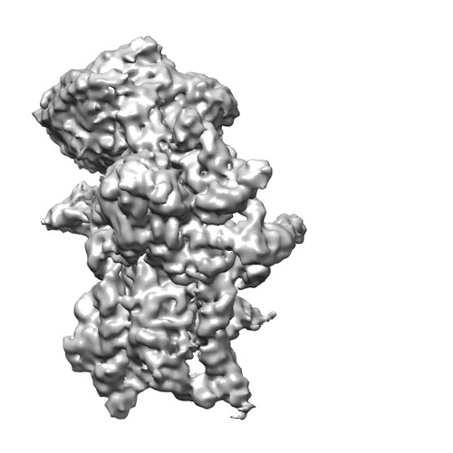 EMD-2450-40S-IRES-DHX29-500X500.jpg EMD-2450-40S-IRES-DHX29-500X500.jpg | 94.3 KB | ||
| アーカイブディレクトリ |  http://ftp.pdbj.org/pub/emdb/structures/EMD-2450ftp://ftp.pdbj.org/pub/emdb/structures/EMD-2450 http://ftp.pdbj.org/pub/emdb/structures/EMD-2450ftp://ftp.pdbj.org/pub/emdb/structures/EMD-2450 | HTTPS FTP |
-関連構造データ
-リンク
| EMDBのページ | EMDB (EBI/PDBe) / EMDataResource |
|---|---|
| 「今月の分子」の関連する項目 |

-マップ
| ファイル | ダウンロード / ファイル: emd_2450.map.gz / 形式: CCP4 / 大きさ: 39.7 MB / タイプ: IMAGE STORED AS FLOATING POINT NUMBER (4 BYTES) | ||||||||||||||||||||||||||||||||||||||||||||||||||||||||||||||||||||
|---|---|---|---|---|---|---|---|---|---|---|---|---|---|---|---|---|---|---|---|---|---|---|---|---|---|---|---|---|---|---|---|---|---|---|---|---|---|---|---|---|---|---|---|---|---|---|---|---|---|---|---|---|---|---|---|---|---|---|---|---|---|---|---|---|---|---|---|---|---|
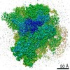
| 注釈 | Reconstruction of a mutant Classical Swine Fever Virus IRES bound to the Rabbit 40S subunit and DHX29. | ||||||||||||||||||||||||||||||||||||||||||||||||||||||||||||||||||||
| 投影像・断面図 | 画像のコントロール
画像は Spider により作成 | ||||||||||||||||||||||||||||||||||||||||||||||||||||||||||||||||||||
| ボクセルのサイズ | X=Y=Z: 2.245 Å | ||||||||||||||||||||||||||||||||||||||||||||||||||||||||||||||||||||
| 密度 |
| ||||||||||||||||||||||||||||||||||||||||||||||||||||||||||||||||||||
| 対称性 | 空間群: 1 | ||||||||||||||||||||||||||||||||||||||||||||||||||||||||||||||||||||
| 詳細 | EMDB XML:
CCP4マップ ヘッダ情報:
| ||||||||||||||||||||||||||||||||||||||||||||||||||||||||||||||||||||
 Z (Sec.)
 Y (Row.)
 X (Col.)


-添付データ
- 試料の構成要素
-全体 : CSFV IRES truncated from domain II, in complex with the Rabbit sm...
| 全体 | 名称: CSFV IRES truncated from domain II, in complex with the Rabbit small ribosomal 40S subunit and to DHX29 |
|---|---|
| 要素 |
|
-超分子 #1000: CSFV IRES truncated from domain II, in complex with the Rabbit sm...
| 超分子 | 名称: CSFV IRES truncated from domain II, in complex with the Rabbit small ribosomal 40S subunit and to DHX29 タイプ: sample / ID: 1000 / 集合状態: one 40S, one CSFV IRES and one DHX29 / Number unique components: 3 |
|---|---|
| 分子量 | 理論値: 1.75 MDa |
-超分子 #1: eukaryotic small ribosmal subunit
| 超分子 | 名称: eukaryotic small ribosmal subunit / タイプ: complex / ID: 1 / Name.synonym: 40S subunit / 組換発現: No / Ribosome-details: ribosome-eukaryote: SSU 40S, SSU RNA 18S |
|---|---|
| 由来(天然) | 生物種: |
| 分子量 | 理論値: 1.5 MDa |
-分子 #1: Internal Ribosomal Entry Site
| 分子 | 名称: Internal Ribosomal Entry Site / タイプ: rna / ID: 1 / Name.synonym: IRES / 分類: OTHER / Structure: DOUBLE HELIX / Synthetic?: No |
|---|---|
| 由来(天然) | 生物種: Classical swine fever virus (豚コレラウイルス) 別称: CSFV |
| 分子量 | 理論値: 100 KDa |
| 配列 | 文字列: GACUAGCCGU AGUGGCGAGC UCCCUGGGUG GUCUAAGUCC UGAGUACAGG ACAGUCGUCA GUAGUUCGAC GUGAGCACUA GCCCACCUCG AGAUGCUACG UGGACGAGGG CAUGCCCAAG ACACACCUUA ACCCUGGCGG GGGUCGCUAG GGUGAAAUCA CAUUAUGUGA ...文字列: GACUAGCCGU AGUGGCGAGC UCCCUGGGUG GUCUAAGUCC UGAGUACAGG ACAGUCGUCA GUAGUUCGAC GUGAGCACUA GCCCACCUCG AGAUGCUACG UGGACGAGGG CAUGCCCAAG ACACACCUUA ACCCUGGCGG GGGUCGCUAG GGUGAAAUCA CAUUAUGUGA UGGGGGUACG ACCUGAUAGG GUGCUGCAGA GGCCCACUAG CAGGCUAGUA UAAAAAUCUC UGC |
-分子 #2: DHX29
| 分子 | 名称: DHX29 / タイプ: protein_or_peptide / ID: 2 / コピー数: 1 / 集合状態: Monomer / 組換発現: Yes |
|---|---|
| 由来(天然) | 生物種: Homo sapiens (ヒト) / 別称: Human |
| 分子量 | 理論値: 150 KDa |
| 組換発現 | 生物種:  |
-実験情報
-構造解析
| 手法 | クライオ電子顕微鏡法 |
|---|---|
 解析 解析 | 単粒子再構成法 |
| 試料の集合状態 | particle |
-試料調製
| 濃度 | 0.105 mg/mL |
|---|---|
| 緩衝液 | pH: 7.5 詳細: 20 mM Tris, 75 mM KCl, 5 mM Mg, 2 mM DTT and 0.25 mM spermidine |
| グリッド | 詳細: 300 mesh copper/molybdenum holey carbon-coated Quantifoil 2/4 grid (Quantifoil Micro Tools GmbH) containing an additional continuous thin layer of carbon |
| 凍結 | 凍結剤: ETHANE / チャンバー内湿度: 100 % / チャンバー内温度: 120 K / 装置: FEI VITROBOT MARK II 手法: 30 seconds waiting after sample deposition on the grid, blotting for seconds before plunging |
- 電子顕微鏡法
| 顕微鏡 | FEI TECNAI F20 |
|---|---|
| 温度 | 平均: 110 K |
| 日付 | 2013年2月1日 |
| 撮影 | カテゴリ: CCD フィルム・検出器のモデル: GATAN ULTRASCAN 4000 (4k x 4k) 実像数: 12000 / 平均電子線量: 12 e/Å2 / ビット/ピクセル: 32 |
| 電子線 | 加速電圧: 120 kV / 電子線源:  FIELD EMISSION GUN FIELD EMISSION GUN |
| 電子光学系 | 倍率(補正後): 51570 / 照射モード: FLOOD BEAM / 撮影モード: BRIGHT FIELD / Cs: 2.26 mm / 最大 デフォーカス(公称値): -4.0 µm / 最小 デフォーカス(公称値): -1.0 µm |
| 試料ステージ | 試料ホルダー: Gatan CT3500 side-entry cryo-holder / 試料ホルダーモデル: OTHER |
| 実験機器 |  モデル: Tecnai F20 / 画像提供: FEI Company |
-画像解析
| 詳細 | The particles constituting this map were sorted from a larger dataset using RELION |
|---|---|
| CTF補正 | 詳細: Each particle |
| 最終 再構成 | 想定した対称性 - 点群: C1 (非対称) / アルゴリズム: OTHER / 解像度のタイプ: BY AUTHOR / 解像度: 8.5 Å / 解像度の算出法: OTHER / ソフトウェア - 名称: Spider, Relion / 使用した粒子像数: 72900 |
