National Institutes of Health/National Institute of General Medical Sciences (NIH/NIGMS)
United States
National Science Foundation (NSF, United States)
United States
Citation
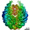
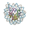
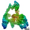
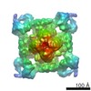
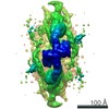
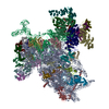
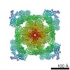
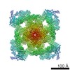
Journal: Nucleic Acids Res / Year: 2021 Title: Structural basis of chromatin regulation by histone variant H2A.Z. Authors: Tyler S Lewis / Vladyslava Sokolova / Harry Jung / Honkit Ng / Dongyan Tan / Abstract: The importance of histone variant H2A.Z in transcription regulation has been well established, yet its mechanism-of-action remains enigmatic. Conflicting evidence exists in support of both an ...The importance of histone variant H2A.Z in transcription regulation has been well established, yet its mechanism-of-action remains enigmatic. Conflicting evidence exists in support of both an activating and a repressive role of H2A.Z in transcription. Here we report cryo-electron microscopy (cryo-EM) structures of nucleosomes and chromatin fibers containing H2A.Z and those containing canonical H2A. The structures show that H2A.Z incorporation results in substantial structural changes in both nucleosome and chromatin fiber. While H2A.Z increases the mobility of DNA terminus in nucleosomes, it simultaneously enables nucleosome arrays to form a more regular and condensed chromatin fiber. We also demonstrated that H2A.Z's ability to enhance nucleosomal DNA mobility is largely attributed to its characteristic shorter C-terminus. Our study provides the structural basis for H2A.Z-mediated chromatin regulation, showing that the increase flexibility of the DNA termini in H2A.Z nucleosomes is central to its dual-functions in chromatin regulation and in transcription.
History
Deposition
Mar 16, 2021
-
Header (metadata) release
Sep 29, 2021
-
Map release
Sep 29, 2021
-
Update
Nov 17, 2021
-
Current status
Nov 17, 2021
Processing site: RCSB / Status: Released
-
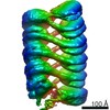
Structure visualization
Movie
Surface view with section colored by density value
A: 303.199 Å / B: 216.879 Å / C: 290.251 Å α=β=γ: 90.0 °
CCP4 map header:
mode
Image stored as Reals
Å/pix. X/Y/Z
1.079
1.079
1.079
M x/y/z
281
201
269
origin x/y/z
0.000
0.000
0.000
length x/y/z
303.199
216.879
290.251
α/β/γ
90.000
90.000
90.000
start NX/NY/NZ
0
0
0
NX/NY/NZ
376
376
376
MAP C/R/S
1
2
3
start NC/NR/NS
58
74
34
NC/NR/NS
281
201
269
D min/max/mean
-0.016
0.078
0.002
-
Supplemental data
-
Sample components
+
Entire : In vitro reconstituted 12x167 bp chromatin
Entire
Name: In vitro reconstituted 12x167 bp chromatin
Components
Complex: In vitro reconstituted 12x167 bp chromatin
Complex: Histone H2A.Z
Protein or peptide: Histone H2A.Z
Complex: Histone H2B
Protein or peptide: Histone H2B
Complex: 12 tandem repeats of 167 bp of 601 Windom nucleosome-positioning sequence
DNA: 12 tandem repeats of 167 bp of 601 Windom nucleosome-positioning sequence
Complex: Histone H3
Protein or peptide: Histone H3
Complex: Histone H4
Protein or peptide: Histone H4
+
Supramolecule #1: In vitro reconstituted 12x167 bp chromatin
Supramolecule
Name: In vitro reconstituted 12x167 bp chromatin / type: complex / ID: 1 / Parent: 0 / Macromolecule list: all Details: EM Map density only contains 8 nucleosomes of the chromatin fiber
Model: Quantifoil R1.2/1.3 / Material: COPPER / Mesh: 400 / Support film - Material: CARBON / Support film - topology: HOLEY / Pretreatment - Type: GLOW DISCHARGE / Pretreatment - Atmosphere: AIR
Vitrification
Cryogen name: ETHANE / Chamber humidity: 100 % / Chamber temperature: 277.15 K / Instrument: FEI VITROBOT MARK IV Details: Sample was absorbed on the grid for thirty seconds, then blotted for five seconds with a blot force zero before plunge freezing..
Details
The sample is crosslinked with 0.1% glutaraldehyde
-
Electron microscopy
Microscope
FEI TITAN KRIOS
Image recording
Film or detector model: GATAN K3 BIOQUANTUM (6k x 4k) / Number grids imaged: 5 / Number real images: 14425 / Average electron dose: 31.0 e/Å2
Electron beam
Acceleration voltage: 300 kV / Electron source: FIELD EMISSION GUN
Type of model: OTHER Details: Ab initio reconstruction in cryoSPARC was used to build the initial model.
Final reconstruction
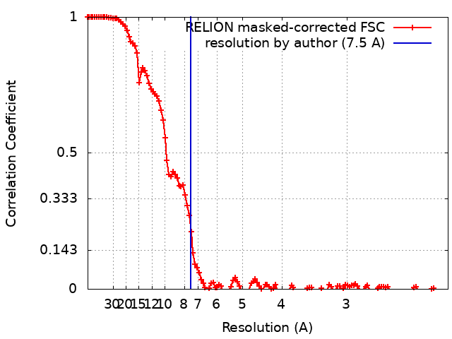
Number classes used: 2 / Applied symmetry - Point group: C1 (asymmetric) / Algorithm: BACK PROJECTION / Resolution.type: BY AUTHOR / Resolution: 7.5 Å / Resolution method: FSC 0.143 CUT-OFF / Software - Name: RELION (ver. 3.1.0) Details: Multi-body refinement based on the consensus refined map was performed to improve the resolution and quantity of the map. Number images used: 111888
Initial angle assignment
Type: MAXIMUM LIKELIHOOD / Software - Name: RELION (ver. 3.1.0)
Final angle assignment
Type: MAXIMUM LIKELIHOOD / Software - Name: RELION (ver. 3.1.0)
Final 3D classification
Number classes: 5 / Software - Name: RELION (ver. 3.1.0)
FSC plot (resolution estimation)
+
About Yorodumi
-
News
-
Feb 9, 2022. New format data for meta-information of EMDB entries
New format data for meta-information of EMDB entries
Version 3 of the EMDB header file is now the official format.
The previous official version 1.9 will be removed from the archive.
In the structure databanks used in Yorodumi, some data are registered as the other names, "COVID-19 virus" and "2019-nCoV". Here are the details of the virus and the list of structure data.
Jan 31, 2019. EMDB accession codes are about to change! (news from PDBe EMDB page)
EMDB accession codes are about to change! (news from PDBe EMDB page)
The allocation of 4 digits for EMDB accession codes will soon come to an end. Whilst these codes will remain in use, new EMDB accession codes will include an additional digit and will expand incrementally as the available range of codes is exhausted. The current 4-digit format prefixed with “EMD-” (i.e. EMD-XXXX) will advance to a 5-digit format (i.e. EMD-XXXXX), and so on. It is currently estimated that the 4-digit codes will be depleted around Spring 2019, at which point the 5-digit format will come into force.
The EM Navigator/Yorodumi systems omit the EMD- prefix.
Related info.:Q: What is EMD? / ID/Accession-code notation in Yorodumi/EM Navigator
Yorodumi is a browser for structure data from EMDB, PDB, SASBDB, etc.
This page is also the successor to EM Navigator detail page, and also detail information page/front-end page for Omokage search.
The word "yorodu" (or yorozu) is an old Japanese word meaning "ten thousand". "mi" (miru) is to see.
Related info.:EMDB / PDB / SASBDB / Comparison of 3 databanks / Yorodumi Search / Aug 31, 2016. New EM Navigator & Yorodumi / Yorodumi Papers / Jmol/JSmol / Function and homology information / Changes in new EM Navigator and Yorodumi
 Movie
Movie Controller


 Open data
 Basic information
 Map data
 Sample
 Function and homology information
 Authors
 United States, 2 items
  Citation
 Structure visualization
 Downloads & links
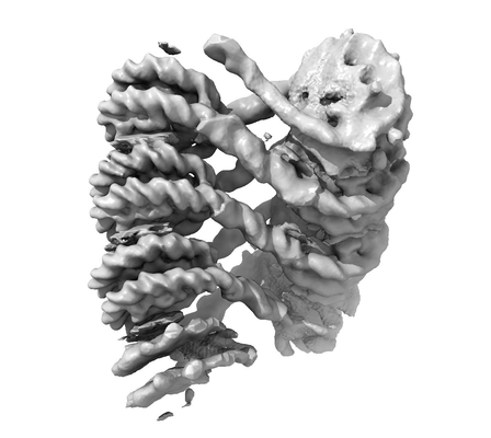 emd_23630.png
 http://ftp.pdbj.org/pub/emdb/structures/EMD-23630
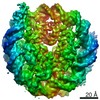


 Z (Sec.)
 Y (Row.)
 X (Col.)


 Sample components
 Processing
 Electron microscopy
 FIELD EMISSION GUN