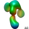
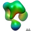
- EMDB-23299: The negative stain EM structure of the DNA Ligase III catalytic c... -
+
Open data
ID or keywords:
Loading...
-
Basic information
Entry
Database: EMDB / ID: EMD-23299
Title
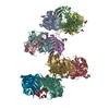
The negative stain EM structure of the DNA Ligase III catalytic core in complex with TDP1.
Map data
DNA LigIII catalytic fragment in complex with TDP1
Sample
Complex: DNA LigIII catalytic core in complex with TDP1
Protein or peptide: Nuclear DNA ligase III
Protein or peptide: Tyrosyl-DNA phosphodiesterase 1
Keywords
DNA repair / protein-protein interactions / DNA BINDING PROTEIN
Function / homology
Function and homology information
DNA ligase III-XRCC1 complex / negative regulation of mitochondrial DNA replication / 3'-tyrosyl-DNA phosphodiesterase activity / DNA ligase activity / DNA ligase (ATP) / DNA ligase (ATP) activity / Strand-asynchronous mitochondrial DNA replication / Hydrolases; Acting on ester bonds; Phosphoric-diester hydrolases / double-strand break repair via alternative nonhomologous end joining / lagging strand elongation ...DNA ligase III-XRCC1 complex / negative regulation of mitochondrial DNA replication / 3'-tyrosyl-DNA phosphodiesterase activity / DNA ligase activity / DNA ligase (ATP) / DNA ligase (ATP) activity / Strand-asynchronous mitochondrial DNA replication / Hydrolases; Acting on ester bonds; Phosphoric-diester hydrolases / double-strand break repair via alternative nonhomologous end joining / lagging strand elongation / HDR through MMEJ (alt-NHEJ) / single strand break repair / Resolution of AP sites via the single-nucleotide replacement pathway / exonuclease activity / mitochondrial DNA repair / DNA biosynthetic process / APEX1-Independent Resolution of AP Sites via the Single Nucleotide Replacement Pathway / base-excision repair, gap-filling / Gap-filling DNA repair synthesis and ligation in GG-NER / mitochondrion organization / Nonhomologous End-Joining (NHEJ) / base-excision repair / double-strand break repair via homologous recombination / Gap-filling DNA repair synthesis and ligation in TC-NER / double-strand break repair / single-stranded DNA binding / double-stranded DNA binding / mitochondrial matrix / cell division / DNA repair / mitochondrion / DNA binding / zinc ion binding / nucleoplasm / ATP binding / nucleus / plasma membrane / cytoplasm Similarity search - Function
DNA ligase 3, BRCT domain / DNA ligase 3 BRCT domain / Tyrosyl-DNA phosphodiesterase I / Tyrosyl-DNA phosphodiesterase / : / DNA ligase, ATP-dependent / DNA ligase, ATP-dependent, N-terminal / DNA ligase, ATP-dependent, N-terminal domain superfamily / DNA ligase N terminus / ATP-dependent DNA ligase AMP-binding site. ...DNA ligase 3, BRCT domain / DNA ligase 3 BRCT domain / Tyrosyl-DNA phosphodiesterase I / Tyrosyl-DNA phosphodiesterase / : / DNA ligase, ATP-dependent / DNA ligase, ATP-dependent, N-terminal / DNA ligase, ATP-dependent, N-terminal domain superfamily / DNA ligase N terminus / ATP-dependent DNA ligase AMP-binding site. / ATP-dependent DNA ligase signature 2. / DNA ligase, ATP-dependent, C-terminal / ATP dependent DNA ligase C terminal region / Zinc finger poly(ADP-ribose) polymerase (PARP)-type signature. / Zinc finger, PARP-type superfamily / Poly(ADP-ribose) polymerase and DNA-Ligase Zn-finger region / Zinc finger poly(ADP-ribose) polymerase (PARP)-type profile. / Poly(ADP-ribose) polymerase and DNA-Ligase Zn-finger region / DNA ligase, ATP-dependent, conserved site / Zinc finger, PARP-type / ATP-dependent DNA ligase family profile. / DNA ligase, ATP-dependent, central / ATP dependent DNA ligase domain / breast cancer carboxy-terminal domain / BRCT domain profile. / BRCT domain / BRCT domain superfamily / Nucleic acid-binding, OB-fold Similarity search - Domain/homology
National Institutes of Health/National Institute of General Medical Sciences (NIH/NIGMS)
R01 ES012512
United States
National Institutes of Health/National Institute of General Medical Sciences (NIH/NIGMS)
R35 CA22043
United States
Natural Sciences and Engineering Research Council (NSERC, Canada)
RGPIN-2015-05776
Canada
Other government
P01 CA92584
United States
Citation
Journal: J Biol Chem / Year: 2021 Title: Direct interaction of DNA repair protein tyrosyl DNA phosphodiesterase 1 and the DNA ligase III catalytic domain is regulated by phosphorylation of its flexible N-terminus. Authors: Ishtiaque Rashid / Michal Hammel / Aleksandr Sverzhinsky / Miaw-Sheue Tsai / John M Pascal / John A Tainer / Alan E Tomkinson / Abstract: Tyrosyl DNA phosphodiesterase 1 (TDP1) and DNA Ligase IIIα (LigIIIα) are key enzymes in single-strand break (SSB) repair. TDP1 removes 3'-tyrosine residues remaining after degradation of DNA ...Tyrosyl DNA phosphodiesterase 1 (TDP1) and DNA Ligase IIIα (LigIIIα) are key enzymes in single-strand break (SSB) repair. TDP1 removes 3'-tyrosine residues remaining after degradation of DNA topoisomerase (TOP) 1 cleavage complexes trapped by either DNA lesions or TOP1 inhibitors. It is not known how TDP1 is linked to subsequent processing and LigIIIα-catalyzed joining of the SSB. Here we define a direct interaction between the TDP1 catalytic domain and the LigIII DNA-binding domain (DBD) regulated by conformational changes in the unstructured TDP1 N-terminal region induced by phosphorylation and/or alterations in amino acid sequence. Full-length and N-terminally truncated TDP1 are more effective at correcting SSB repair defects in TDP1 null cells compared with full-length TDP1 with amino acid substitutions of an N-terminal serine residue phosphorylated in response to DNA damage. TDP1 forms a stable complex with LigIII, as well as full-length LigIIIα alone or in complex with the DNA repair scaffold protein XRCC1. Small-angle X-ray scattering and negative stain electron microscopy combined with mapping of the interacting regions identified a TDP1/LigIIIα compact dimer of heterodimers in which the two LigIII catalytic cores are positioned in the center, whereas the two TDP1 molecules are located at the edges of the core complex flanked by highly flexible regions that can interact with other repair proteins and SSBs. As TDP1and LigIIIα together repair adducts caused by TOP1 cancer chemotherapy inhibitors, the defined interaction architecture and regulation of this enzyme complex provide insights into a key repair pathway in nonmalignant and cancer cells.
History
Deposition
Jan 15, 2021
-
Header (metadata) release
Jul 14, 2021
-
Map release
Jul 14, 2021
-
Update
Nov 29, 2023
-
Current status
Nov 29, 2023
Processing site: RCSB / Status: Released
-
Structure visualization
Movie
Surface view with section colored by density value
In the structure databanks used in Yorodumi, some data are registered as the other names, "COVID-19 virus" and "2019-nCoV". Here are the details of the virus and the list of structure data.
Jan 31, 2019. EMDB accession codes are about to change! (news from PDBe EMDB page)
EMDB accession codes are about to change! (news from PDBe EMDB page)
The allocation of 4 digits for EMDB accession codes will soon come to an end. Whilst these codes will remain in use, new EMDB accession codes will include an additional digit and will expand incrementally as the available range of codes is exhausted. The current 4-digit format prefixed with “EMD-” (i.e. EMD-XXXX) will advance to a 5-digit format (i.e. EMD-XXXXX), and so on. It is currently estimated that the 4-digit codes will be depleted around Spring 2019, at which point the 5-digit format will come into force.
The EM Navigator/Yorodumi systems omit the EMD- prefix.
Related info.:Q: What is EMD? / ID/Accession-code notation in Yorodumi/EM Navigator
Yorodumi is a browser for structure data from EMDB, PDB, SASBDB, etc.
This page is also the successor to EM Navigator detail page, and also detail information page/front-end page for Omokage search.
The word "yorodu" (or yorozu) is an old Japanese word meaning "ten thousand". "mi" (miru) is to see.
Related info.:EMDB / PDB / SASBDB / Comparison of 3 databanks / Yorodumi Search / Aug 31, 2016. New EM Navigator & Yorodumi / Yorodumi Papers / Jmol/JSmol / Function and homology information / Changes in new EM Navigator and Yorodumi
 Movie
Movie Controller

 Yorodumi
 Open data
 Basic information
 Map data
 Sample
 Keywords
 Function and homology information
 Homo sapiens (human)
 Authors
 United States,
  Canada, 4 items
  Citation
 Structure visualization
 UCSF Chimera
 Downloads & links
 emd_23299.png
 http://ftp.pdbj.org/pub/emdb/structures/EMD-23299


 Z (Sec.)
 Y (Row.)
 X (Col.)


 Sample components
 Spodoptera frugiperda (fall armyworm)
 Processing
 Electron microscopy