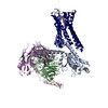
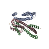
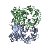
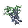
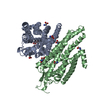
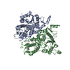
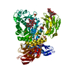
Journal: Nature / Year: 2020 Title: Structure of a D2 dopamine receptor-G-protein complex in a lipid membrane. Authors: Jie Yin / Kuang-Yui M Chen / Mary J Clark / Mahdi Hijazi / Punita Kumari / Xiao-Chen Bai / Roger K Sunahara / Patrick Barth / Daniel M Rosenbaum / Abstract: The D2 dopamine receptor (DRD2) is a therapeutic target for Parkinson's disease and antipsychotic drugs. DRD2 is activated by the endogenous neurotransmitter dopamine and synthetic agonist drugs such ...The D2 dopamine receptor (DRD2) is a therapeutic target for Parkinson's disease and antipsychotic drugs. DRD2 is activated by the endogenous neurotransmitter dopamine and synthetic agonist drugs such as bromocriptine, leading to stimulation of G and inhibition of adenylyl cyclase. Here we used cryo-electron microscopy to elucidate the structure of an agonist-bound activated DRD2-G complex reconstituted into a phospholipid membrane. The extracellular ligand-binding site of DRD2 is remodelled in response to agonist binding, with conformational changes in extracellular loop 2, transmembrane domain 5 (TM5), TM6 and TM7, propagating to opening of the intracellular G-binding site. The DRD2-G structure represents, to our knowledge, the first experimental model of a G-protein-coupled receptor-G-protein complex embedded in a phospholipid bilayer, which serves as a benchmark to validate the interactions seen in previous detergent-bound structures. The structure also reveals interactions that are unique to the membrane-embedded complex, including helix 8 burial in the inner leaflet, ordered lysine and arginine side chains in the membrane interfacial regions, and lipid anchoring of the G protein in the membrane. Our model of the activated DRD2 will help to inform the design of subtype-selective DRD2 ligands for multiple human central nervous system disorders.
History
Deposition
Jan 28, 2020
-
Header (metadata) release
Feb 12, 2020
-
Map release
Jun 17, 2020
-
Update
Jun 17, 2020
-
Current status
Jun 17, 2020
Processing site: RCSB / Status: Released
-
Structure visualization
Movie
Surface view with section colored by density value
In the structure databanks used in Yorodumi, some data are registered as the other names, "COVID-19 virus" and "2019-nCoV". Here are the details of the virus and the list of structure data.
Jan 31, 2019. EMDB accession codes are about to change! (news from PDBe EMDB page)
EMDB accession codes are about to change! (news from PDBe EMDB page)
The allocation of 4 digits for EMDB accession codes will soon come to an end. Whilst these codes will remain in use, new EMDB accession codes will include an additional digit and will expand incrementally as the available range of codes is exhausted. The current 4-digit format prefixed with “EMD-” (i.e. EMD-XXXX) will advance to a 5-digit format (i.e. EMD-XXXXX), and so on. It is currently estimated that the 4-digit codes will be depleted around Spring 2019, at which point the 5-digit format will come into force.
The EM Navigator/Yorodumi systems omit the EMD- prefix.
Related info.:Q: What is EMD? / ID/Accession-code notation in Yorodumi/EM Navigator
Yorodumi is a browser for structure data from EMDB, PDB, SASBDB, etc.
This page is also the successor to EM Navigator detail page, and also detail information page/front-end page for Omokage search.
The word "yorodu" (or yorozu) is an old Japanese word meaning "ten thousand". "mi" (miru) is to see.
Related info.:EMDB / PDB / SASBDB / Comparison of 3 databanks / Yorodumi Search / Aug 31, 2016. New EM Navigator & Yorodumi / Yorodumi Papers / Jmol/JSmol / Function and homology information / Changes in new EM Navigator and Yorodumi
 Movie
Movie Controller

 Yorodumi
 Open data
 Basic information
 Map data
 Sample
 Homo sapiens (human)
 Authors
 United States, 1 items
  Citation
 Structure visualization
 Movie viewer
 Downloads & links
 emd_21245.png
 http://ftp.pdbj.org/pub/emdb/structures/EMD-21245
 Z (Sec.)
 Y (Row.)
 X (Col.)


 Sample components
 Spodoptera frugiperda (fall armyworm)
 Processing
 Electron microscopy
 FIELD EMISSION GUN