Movie
Movie Controller

[English] 日本語
 Yorodumi
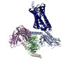
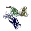
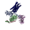
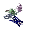
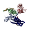
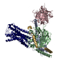
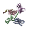
- PDB-6vms: Structure of a D2 dopamine receptor-G-protein complex in a lipid ... -
+ Open data

- Basic information
| Entry | Database: PDB / ID: 6vms | ||||||
|---|---|---|---|---|---|---|---|
| Title | Structure of a D2 dopamine receptor-G-protein complex in a lipid membrane | ||||||
 Components Components |
| ||||||
 Keywords Keywords | SIGNALING PROTEIN / Dopamine / Dopamine receptor / GPCR / G protein / Parkinson's disease | ||||||
| Function / homology |  Function and homology information Function and homology informationnegative regulation of dopamine receptor signaling pathway / positive regulation of dopamine uptake involved in synaptic transmission / negative regulation of dephosphorylation / positive regulation of glial cell-derived neurotrophic factor production / nervous system process involved in regulation of systemic arterial blood pressure / acid secretion / regulation of synapse structural plasticity / dopamine neurotransmitter receptor activity, coupled via Gi/Go / response to histamine / negative regulation of circadian sleep/wake cycle, sleep ...negative regulation of dopamine receptor signaling pathway / positive regulation of dopamine uptake involved in synaptic transmission / negative regulation of dephosphorylation / positive regulation of glial cell-derived neurotrophic factor production / nervous system process involved in regulation of systemic arterial blood pressure / acid secretion / regulation of synapse structural plasticity / dopamine neurotransmitter receptor activity, coupled via Gi/Go / response to histamine / negative regulation of circadian sleep/wake cycle, sleep / regulation of locomotion involved in locomotory behavior / neuron-neuron synaptic transmission / adenylate cyclase regulator activity / Extra-nuclear estrogen signaling / Adenylate cyclase inhibitory pathway / negative regulation of cellular response to hypoxia / adenylate cyclase-inhibiting dopamine receptor signaling pathway / response to inactivity / regulation of potassium ion transport / orbitofrontal cortex development / adenohypophysis development / negative regulation of dopamine secretion / cerebral cortex GABAergic interneuron migration / negative regulation of neuron migration / hyaloid vascular plexus regression / Dopamine receptors / branching morphogenesis of a nerve / regulation of dopamine uptake involved in synaptic transmission / dopamine binding / positive regulation of growth hormone secretion / phospholipase C-activating dopamine receptor signaling pathway / heterotrimeric G-protein binding / peristalsis / G protein-coupled receptor complex / beta-arrestin-dependent dopamine receptor signaling pathway / grooming behavior / positive regulation of renal sodium excretion / drinking behavior / striatum development / auditory behavior / positive regulation of G protein-coupled receptor signaling pathway / dopaminergic synapse / behavioral response to ethanol / positive regulation of multicellular organism growth / non-motile cilium / negative regulation of synaptic transmission / G protein-coupled receptor internalization / Adrenaline,noradrenaline inhibits insulin secretion / ADP signalling through P2Y purinoceptor 12 / response to iron ion / GTPase activating protein binding / G alpha (i) signalling events / adult walking behavior / negative regulation of synaptic transmission, glutamatergic / arachidonate secretion / positive regulation of urine volume / positive regulation of neuroblast proliferation / ciliary membrane / neurotransmitter receptor localization to postsynaptic specialization membrane / positive regulation of cytokinesis / regulation of synaptic transmission, GABAergic / response to morphine / negative regulation of cytosolic calcium ion concentration / dopamine metabolic process / temperature homeostasis / pigmentation / dopamine uptake involved in synaptic transmission / regulation of dopamine secretion / neuroblast proliferation / positive regulation of receptor internalization / lateral plasma membrane / associative learning / negative regulation of protein secretion / response to light stimulus / cellular response to ethanol / potassium channel regulator activity / endocytic vesicle / G-protein alpha-subunit binding / response to axon injury / viral release from host cell by cytolysis / sperm flagellum / postsynaptic modulation of chemical synaptic transmission / prepulse inhibition / long-term memory / regulation of sodium ion transport / cellular response to retinoic acid / negative regulation of blood pressure / peptidoglycan catabolic process / release of sequestered calcium ion into cytosol / behavioral response to cocaine / adenylate cyclase inhibitor activity / positive regulation of protein localization to cell cortex / T cell migration / synapse assembly / axonogenesis / D2 dopamine receptor binding / epithelial cell proliferation / negative regulation of innate immune response / regulation of heart rate / adenylate cyclase-inhibiting serotonin receptor signaling pathway Similarity search - Function | ||||||
| Biological species |   Homo sapiens (human) Homo sapiens (human) Enterobacteria phage T4 (virus) Enterobacteria phage T4 (virus) | ||||||
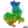
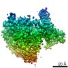
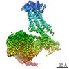
| Method | ELECTRON MICROSCOPY / single particle reconstruction / cryo EM / Resolution: 3.8 Å | ||||||
 Authors Authors | Yin, J. / Chen, K.M. / Clark, M.J. / Hijazi, M. / Kumari, P. / Bai, X. / Sunahara, R.K. / Barth, P. / Rosenbaum, D.M. | ||||||
| Funding support |  United States, 1items United States, 1items
| ||||||
 Citation Citation | Journal: Nature / Year: 2020 Title: Structure of a D2 dopamine receptor-G-protein complex in a lipid membrane. Authors: Jie Yin / Kuang-Yui M Chen / Mary J Clark / Mahdi Hijazi / Punita Kumari / Xiao-Chen Bai / Roger K Sunahara / Patrick Barth / Daniel M Rosenbaum /  Abstract: The D2 dopamine receptor (DRD2) is a therapeutic target for Parkinson's disease and antipsychotic drugs. DRD2 is activated by the endogenous neurotransmitter dopamine and synthetic agonist drugs such ...The D2 dopamine receptor (DRD2) is a therapeutic target for Parkinson's disease and antipsychotic drugs. DRD2 is activated by the endogenous neurotransmitter dopamine and synthetic agonist drugs such as bromocriptine, leading to stimulation of G and inhibition of adenylyl cyclase. Here we used cryo-electron microscopy to elucidate the structure of an agonist-bound activated DRD2-G complex reconstituted into a phospholipid membrane. The extracellular ligand-binding site of DRD2 is remodelled in response to agonist binding, with conformational changes in extracellular loop 2, transmembrane domain 5 (TM5), TM6 and TM7, propagating to opening of the intracellular G-binding site. The DRD2-G structure represents, to our knowledge, the first experimental model of a G-protein-coupled receptor-G-protein complex embedded in a phospholipid bilayer, which serves as a benchmark to validate the interactions seen in previous detergent-bound structures. The structure also reveals interactions that are unique to the membrane-embedded complex, including helix 8 burial in the inner leaflet, ordered lysine and arginine side chains in the membrane interfacial regions, and lipid anchoring of the G protein in the membrane. Our model of the activated DRD2 will help to inform the design of subtype-selective DRD2 ligands for multiple human central nervous system disorders. | ||||||
| History |
|
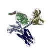
- Structure visualization
| Movie |
Movie viewer |
|---|---|
| Structure viewer | Molecule: MolmilJmol/JSmol |
- Downloads & links
-Download
| PDBx/mmCIF format | 6vms.cif.gz | 219.7 KB | Display | PDBx/mmCIF format |
|---|---|---|---|---|
| PDB format | pdb6vms.ent.gz | 167.5 KB | Display | PDB format |
| PDBx/mmJSON format | 6vms.json.gz | Tree view | PDBx/mmJSON format | |
| Others |  Other downloads Other downloads |
-Validation report
| Arichive directory | https://data.pdbj.org/pub/pdb/validation_reports/vm/6vmsftp://data.pdbj.org/pub/pdb/validation_reports/vm/6vms | HTTPS FTP |
|---|
-Related structure data
| Related structure data |  21243MC M: map data used to model this data C: citing same article ( |
|---|---|
| Similar structure data |

-Links
 PDBj
PDBj


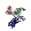
- Assembly
| Deposited unit | 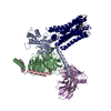
|
|---|---|
| 1 |
|
-Components
-Guanine nucleotide-binding protein ... , 3 types, 3 molecules ABC
| #1: Protein | Mass: 40382.047 Da / Num. of mol.: 1 Source method: isolated from a genetically manipulated source Source: (gene. exp.)   Spodoptera frugiperda (fall armyworm) / References: UniProt: P10824 Spodoptera frugiperda (fall armyworm) / References: UniProt: P10824 |
|---|---|
| #2: Protein | Mass: 37416.930 Da / Num. of mol.: 1 Source method: isolated from a genetically manipulated source Source: (gene. exp.) Homo sapiens (human) / Gene: GNB1 / Production host: Spodoptera frugiperda (fall armyworm) / References: UniProt: P62873 |
| #3: Protein | Mass: 9137.474 Da / Num. of mol.: 1 Source method: isolated from a genetically manipulated source Source: (gene. exp.) Homo sapiens (human) / Gene: GNG2 / Production host: Spodoptera frugiperda (fall armyworm) / References: UniProt: P59768 |
-Antibody / Protein / Non-polymers , 3 types, 3 molecules ER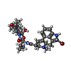
| #4: Antibody | Mass: 27784.896 Da / Num. of mol.: 1 Source method: isolated from a genetically manipulated source Source: (gene. exp.) Homo sapiens (human) / Production host: Spodoptera frugiperda (fall armyworm) |
|---|---|
| #5: Protein | Mass: 51384.059 Da / Num. of mol.: 1 Source method: isolated from a genetically manipulated source Source: (gene. exp.) Enterobacteria phage T4 (virus), (gene. exp.) Homo sapiens (human)Gene: e, T4Tp126, DRD2 / Production host: Spodoptera frugiperda (fall armyworm) / References: UniProt: D9IEF7, UniProt: P14416, lysozyme |
| #6: Chemical | ChemComp-08Y /  Mass: 654.594 Da / Num. of mol.: 1 / Source method: obtained synthetically / Formula: C32H40BrN5O5 / Feature type: SUBJECT OF INVESTIGATION / Comment: agonist*YM Mass: 654.594 Da / Num. of mol.: 1 / Source method: obtained synthetically / Formula: C32H40BrN5O5 / Feature type: SUBJECT OF INVESTIGATION / Comment: agonist*YM |
-Details
| Has ligand of interest | Y |
|---|---|
| Has protein modification | Y |
-Experimental details
-Experiment
| Experiment | Method: ELECTRON MICROSCOPY |
|---|---|
| EM experiment | Aggregation state: PARTICLE / 3D reconstruction method: single particle reconstruction |
- Sample preparation
| Component | Name: D2 dopamine receptor-G protein complex / Type: COMPLEX / Entity ID: #1-#5 / Source: RECOMBINANT |
|---|---|
| Source (natural) | Organism: Homo sapiens (human) |
| Source (recombinant) | Organism: Spodoptera frugiperda (fall armyworm) |
| Buffer solution | pH: 7.4 |
| Specimen | Embedding applied: NO / Shadowing applied: NO / Staining applied: NO / Vitrification applied: YES |
| Vitrification | Cryogen name: ETHANE |
- Electron microscopy imaging
| Experimental equipment |  Model: Titan Krios / Image courtesy: FEI Company |
|---|---|
| Microscopy | Model: FEI TITAN KRIOS |
| Electron gun | Electron source:  FIELD EMISSION GUN / Accelerating voltage: 300 kV / Illumination mode: FLOOD BEAM FIELD EMISSION GUN / Accelerating voltage: 300 kV / Illumination mode: FLOOD BEAM |
| Electron lens | Mode: BRIGHT FIELD |
| Image recording | Electron dose: 64 e/Å2 / Film or detector model: GATAN K3 BIOQUANTUM (6k x 4k) |
- Processing
| EM software |
| ||||||||||||||||||||||||||||||||
|---|---|---|---|---|---|---|---|---|---|---|---|---|---|---|---|---|---|---|---|---|---|---|---|---|---|---|---|---|---|---|---|---|---|
| CTF correction | Type: PHASE FLIPPING AND AMPLITUDE CORRECTION | ||||||||||||||||||||||||||||||||
| Symmetry | Point symmetry: C1 (asymmetric) | ||||||||||||||||||||||||||||||||
| 3D reconstruction | Resolution: 3.8 Å / Resolution method: FSC 0.143 CUT-OFF / Num. of particles: 783984 / Symmetry type: POINT | ||||||||||||||||||||||||||||||||
| Atomic model building | PDB-ID: 6DDE Accession code: 6DDE / Source name: PDB / Type: experimental model |