 Movie
Movie Controller

[English] 日本語
 Yorodumi
- PDB-3zev: Structure of Thermostable Agonist-bound Neurotensin Receptor 1 Mu... -
+ Open data

- Basic information
| Entry | Database: PDB / ID: 3zev | ||||||
|---|---|---|---|---|---|---|---|
| Title | Structure of Thermostable Agonist-bound Neurotensin Receptor 1 Mutant without Lysozyme Fusion | ||||||
 Components Components |
| ||||||
 Keywords Keywords | SIGNALING PROTEIN / MEMBRANE PROTEIN | ||||||
| Function / homology |  Function and homology information Function and homology informationregulation of locomotion involved in locomotory behavior / Peptide ligand-binding receptors / neuropeptide receptor binding / G protein-coupled neurotensin receptor activity / inositol phosphate catabolic process / symmetric synapse / positive regulation of locomotion / regulation of inositol trisphosphate biosynthetic process / response to antipsychotic drug / positive regulation of gamma-aminobutyric acid secretion ...regulation of locomotion involved in locomotory behavior / Peptide ligand-binding receptors / neuropeptide receptor binding / G protein-coupled neurotensin receptor activity / inositol phosphate catabolic process / symmetric synapse / positive regulation of locomotion / regulation of inositol trisphosphate biosynthetic process / response to antipsychotic drug / positive regulation of gamma-aminobutyric acid secretion / D-aspartate import across plasma membrane / neuron spine / neuropeptide hormone activity / positive regulation of arachidonate secretion / vocalization behavior / L-glutamate import across plasma membrane / regulation of behavioral fear response / cAMP biosynthetic process / regulation of respiratory gaseous exchange / hyperosmotic response / negative regulation of systemic arterial blood pressure / G alpha (q) signalling events / digestive tract development / positive regulation of inhibitory postsynaptic potential / negative regulation of release of sequestered calcium ion into cytosol / cellular response to lithium ion / response to mineralocorticoid / positive regulation of glutamate secretion / response to corticosterone / response to food / regulation of membrane depolarization / response to lipid / temperature homeostasis / positive regulation of inositol phosphate biosynthetic process / detection of temperature stimulus involved in sensory perception of pain / response to stress / associative learning / cellular response to dexamethasone stimulus / response to axon injury / conditioned place preference / transport vesicle / blood vessel diameter maintenance / axon terminus / positive regulation of release of sequestered calcium ion into cytosol / dendritic shaft / learning / response to amphetamine / adult locomotory behavior / response to cocaine / liver development / neuropeptide signaling pathway / cellular response to nerve growth factor stimulus / visual learning / cytoplasmic side of plasma membrane / terminal bouton / response to estradiol / dendritic spine / perikaryon / positive regulation of phosphatidylinositol 3-kinase/protein kinase B signal transduction / positive regulation of apoptotic process / membrane raft / receptor ligand activity / negative regulation of gene expression / axon / neuronal cell body / positive regulation of gene expression / dendrite / negative regulation of apoptotic process / protein-containing complex binding / cell surface / extracellular region / identical protein binding / plasma membrane Similarity search - Function | ||||||
| Biological species |  | ||||||
| Method |  X-RAY DIFFRACTION / SYNCHROTRON / OTHER / Resolution: 3 Å X-RAY DIFFRACTION / SYNCHROTRON / OTHER / Resolution: 3 Å | ||||||
 Authors Authors | Egloff, P. / Hillenbrand, M. / Schlinkmann, K.M. / Batyuk, A. / Mittl, P. / Plueckthun, A. | ||||||
 Citation Citation | Journal: Proc.Natl.Acad.Sci.USA / Year: 2014 Title: Structure of Signaling-Competent Neurotensin Receptor 1 Obtained by Directed Evolution in Escherichia Coli Authors: Egloff, P. / Hillenbrand, M. / Klenk, C. / Batyuk, A. / Heine, P. / Balada, S. / Schlinkmann, K.M. / Scott, D.J. / Schuetz, M. / Plueckthun, A. | ||||||
| History |
|
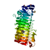
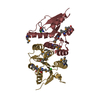
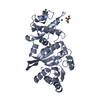
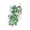
- Structure visualization
| Structure viewer | Molecule: MolmilJmol/JSmol |
|---|
- Downloads & links
-Download
| PDBx/mmCIF format | 3zev.cif.gz | 259.4 KB | Display | PDBx/mmCIF format |
|---|---|---|---|---|
| PDB format | pdb3zev.ent.gz | 213.9 KB | Display | PDB format |
| PDBx/mmJSON format | 3zev.json.gz | Tree view | PDBx/mmJSON format | |
| Others |  Other downloads Other downloads |
-Validation report
| Arichive directory | https://data.pdbj.org/pub/pdb/validation_reports/ze/3zevftp://data.pdbj.org/pub/pdb/validation_reports/ze/3zev | HTTPS FTP |
|---|
-Related structure data


-Links
 PDBj
PDBj


- Assembly
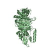
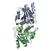
| Deposited unit | 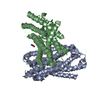
| ||||||||
|---|---|---|---|---|---|---|---|---|---|
| 1 | 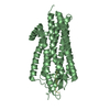
| ||||||||
| 2 | 
| ||||||||
| Unit cell |
| ||||||||
| Noncrystallographic symmetry (NCS) | NCS oper: (Code: given Matrix: (-0.51, 0.1456, 0.8478), Vector: |
-Components
| #1: Protein | Mass: 37607.113 Da / Num. of mol.: 2 / Mutation: YES Source method: isolated from a genetically manipulated source Details: THERMOSTABLE MUTANT / Source: (gene. exp.)  #2: Protein/peptide | Mass: 933.111 Da / Num. of mol.: 2 / Fragment: C-TERMINUS, RESIDUES 157-162 Source method: isolated from a genetically manipulated source Details: RESIDUES 8-13 CORRESPOND TO NEUROTENSIN C-TERMINUS. RESIDUES 6-7 DO NOT CORRESPOND TO NEUROTENSIN SEQUENCE Source: (gene. exp.) #3: Chemical | ChemComp-GLY /   Type: peptide linking / Mass: 75.067 Da / Num. of mol.: 5 / Source method: obtained synthetically / Formula: C2H5NO2 Type: peptide linking / Mass: 75.067 Da / Num. of mol.: 5 / Source method: obtained synthetically / Formula: C2H5NO2Has protein modification | Y | |
|---|
-Experimental details
-Experiment
| Experiment | Method: X-RAY DIFFRACTION / Number of used crystals: 1 |
|---|
- Sample preparation
| Crystal | Density Matthews: 3.68 Å3/Da / Density % sol: 66.54 % / Description: NONE |
|---|---|
| Crystal grow | pH: 9.4 Details: 1.28% (W/V) NONYL-GLUCOSIDE, 0.5% (W/V) DECYL-GLUCOSIDE, 0.01% (W/V) DODECYL-GLUCOSIDE, 0.1% (W/V) CHOLESTERYLHEMISUCCINATE, 10MM HEPES PH 8, 1.15 MM NACL, 2 MM DTT, 100 NM NTI, 26% (V/V) ...Details: 1.28% (W/V) NONYL-GLUCOSIDE, 0.5% (W/V) DECYL-GLUCOSIDE, 0.01% (W/V) DODECYL-GLUCOSIDE, 0.1% (W/V) CHOLESTERYLHEMISUCCINATE, 10MM HEPES PH 8, 1.15 MM NACL, 2 MM DTT, 100 NM NTI, 26% (V/V) PEG 600, 50 MM GLYCINE PH 9.4 |
-Data collection
| Diffraction | Mean temperature: 100 K |
|---|---|
| Diffraction source | Source: SYNCHROTRON / Site: SLS  / Beamline: X06SA / Wavelength: 1 / Beamline: X06SA / Wavelength: 1 |
| Detector | Type: DECTRIS PILATUS 6M / Detector: PIXEL |
| Radiation | Protocol: SINGLE WAVELENGTH / Monochromatic (M) / Laue (L): M / Scattering type: x-ray |
| Radiation wavelength | Wavelength: 1 Å / Relative weight: 1 |
| Reflection | Resolution: 3→50 Å / Num. obs: 22942 / % possible obs: 99.8 % / Observed criterion σ(I): 0.72 / Redundancy: 6.9 % / Biso Wilson estimate: 115.97 Å2 / Rmerge(I) obs: 0.01 / Net I/σ(I): 8.68 |
- Processing
| Software |
| |||||||||||||||||||||||||||||||||||||||||||||||||||||||||||||||||||||||||||
|---|---|---|---|---|---|---|---|---|---|---|---|---|---|---|---|---|---|---|---|---|---|---|---|---|---|---|---|---|---|---|---|---|---|---|---|---|---|---|---|---|---|---|---|---|---|---|---|---|---|---|---|---|---|---|---|---|---|---|---|---|---|---|---|---|---|---|---|---|---|---|---|---|---|---|---|---|
| Refinement | Method to determine structure: OTHER Starting model: NONE Resolution: 3→19.935 Å / SU ML: 0.39 / σ(F): 1.33 / Phase error: 32.74 / Stereochemistry target values: ML
| |||||||||||||||||||||||||||||||||||||||||||||||||||||||||||||||||||||||||||
| Solvent computation | Shrinkage radii: 0.9 Å / VDW probe radii: 1.11 Å / Solvent model: FLAT BULK SOLVENT MODEL | |||||||||||||||||||||||||||||||||||||||||||||||||||||||||||||||||||||||||||
| Displacement parameters | Biso mean: 125.7 Å2 | |||||||||||||||||||||||||||||||||||||||||||||||||||||||||||||||||||||||||||
| Refinement step | Cycle: LAST / Resolution: 3→19.935 Å
| |||||||||||||||||||||||||||||||||||||||||||||||||||||||||||||||||||||||||||
| Refine LS restraints |
| |||||||||||||||||||||||||||||||||||||||||||||||||||||||||||||||||||||||||||
| LS refinement shell |
| |||||||||||||||||||||||||||||||||||||||||||||||||||||||||||||||||||||||||||
| Refinement TLS params. | Method: refined / Refine-ID: X-RAY DIFFRACTION
| |||||||||||||||||||||||||||||||||||||||||||||||||||||||||||||||||||||||||||
| Refinement TLS group |
|
