 Movie
Movie Controller

[English] 日本語
 Yorodumi
- PDB-2ejb: Crystal Structure Of Phenylacrylic Acid Decarboxylase from Aquife... -
+ Open data

- Basic information
| Entry | Database: PDB / ID: 2ejb | ||||||
|---|---|---|---|---|---|---|---|
| Title | Crystal Structure Of Phenylacrylic Acid Decarboxylase from Aquifex aeolicus | ||||||
 Components Components | Probable aromatic acid decarboxylase | ||||||
 Keywords Keywords | LYASE / Phenylacrylic Acid Decarboxylase / Structural Genomics / NPPSFA / National Project on Protein Structural and Functional Analyses / RIKEN Structural Genomics/Proteomics Initiative / RSGI | ||||||
| Function / homology |  Function and homology information Function and homology informationflavin prenyltransferase / flavin prenyltransferase activity / carboxy-lyase activity Similarity search - Function | ||||||
| Biological species |   Aquifex aeolicus (bacteria) Aquifex aeolicus (bacteria) | ||||||
| Method |  X-RAY DIFFRACTION / MOLECULAR REPLACEMENT / Resolution: 2.15 Å X-RAY DIFFRACTION / MOLECULAR REPLACEMENT / Resolution: 2.15 Å | ||||||
 Authors Authors | Bagautdinov, B. / Kunishima, N. / RIKEN Structural Genomics/Proteomics Initiative (RSGI) | ||||||
 Citation Citation | Journal: To be Published Title: Crystal Structure Of Phenylacrylic Acid Decarboxylase from Aquifex aeolicus Authors: Bagautdinov, B. / Kunishima, N. | ||||||
| History |
|
- Structure visualization
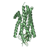
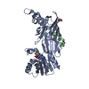
| Structure viewer | Molecule: MolmilJmol/JSmol |
|---|
- Downloads & links
-Download
| PDBx/mmCIF format | 2ejb.cif.gz | 49.4 KB | Display | PDBx/mmCIF format |
|---|---|---|---|---|
| PDB format | pdb2ejb.ent.gz | 35.3 KB | Display | PDB format |
| PDBx/mmJSON format | 2ejb.json.gz | Tree view | PDBx/mmJSON format | |
| Others |  Other downloads Other downloads |
-Validation report
| Arichive directory | https://data.pdbj.org/pub/pdb/validation_reports/ej/2ejbftp://data.pdbj.org/pub/pdb/validation_reports/ej/2ejb | HTTPS FTP |
|---|
-Related structure data
| Related structure data |  1sbzS S: Starting model for refinement |
|---|---|
| Similar structure data | |
| Other databases |


-Links
 PDBj
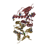
PDBj- Assembly
| Deposited unit | 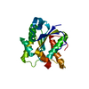
| |||||||||||||||
|---|---|---|---|---|---|---|---|---|---|---|---|---|---|---|---|---|
| 1 | 
| |||||||||||||||
| 2 | x 12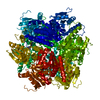
| |||||||||||||||
| Unit cell |
| |||||||||||||||
| Components on special symmetry positions |
|
-Components
| #1: Protein | Mass: 21285.934 Da / Num. of mol.: 1 Source method: isolated from a genetically manipulated source Source: (gene. exp.) Aquifex aeolicus (bacteria) / Plasmid: pET11a / Production host: References: UniProt: O66811, Lyases; Carbon-carbon lyases; Carboxy-lyases |
|---|---|
| #2: Water | ChemComp-HOH /  Mass: 18.015 Da / Num. of mol.: 79 / Source method: isolated from a natural source / Formula: H2O Mass: 18.015 Da / Num. of mol.: 79 / Source method: isolated from a natural source / Formula: H2O |
-Experimental details
-Experiment
| Experiment | Method: X-RAY DIFFRACTION / Number of used crystals: 1 |
|---|
- Sample preparation
| Crystal | Density Matthews: 2.2 Å3/Da / Density % sol: 43.99 % |
|---|---|
| Crystal grow | Temperature: 295 K / Method: microbatch / pH: 4.5 Details: 10% PEG 6000, 2M NaCl, pH 4.5, microbatch, temperature 295K |
-Data collection
| Diffraction | Mean temperature: 100 K |
|---|---|
| Diffraction source | Source: ROTATING ANODE / Type: RIGAKU / Wavelength: 1.5418 Å |
| Detector | Type: RIGAKU RAXIS IV / Detector: IMAGE PLATE / Date: Jan 12, 2006 / Details: mirrors |
| Radiation | Monochromator: GRAPHITE / Protocol: SINGLE WAVELENGTH / Monochromatic (M) / Laue (L): M / Scattering type: x-ray |
| Radiation wavelength | Wavelength: 1.5418 Å / Relative weight: 1 |
| Reflection | Resolution: 2.15→32.73 Å / Num. all: 10214 / Num. obs: 9696 / % possible obs: 94.9 % / Observed criterion σ(F): 0 / Observed criterion σ(I): 0 / Redundancy: 3.5 % / Biso Wilson estimate: 44.3 Å2 / Rmerge(I) obs: 0.048 / Rsym value: 0.038 / Net I/σ(I): 36.8 |
| Reflection shell | Resolution: 2.15→2.23 Å / Redundancy: 3 % / Rmerge(I) obs: 0.309 / Mean I/σ(I) obs: 4.8 / Num. unique all: 954 / Rsym value: 0.294 / % possible all: 95.4 |
- Processing
| Software |
| |||||||||||||||||||||||||
|---|---|---|---|---|---|---|---|---|---|---|---|---|---|---|---|---|---|---|---|---|---|---|---|---|---|---|
| Refinement | Method to determine structure: MOLECULAR REPLACEMENT Starting model: PDB ENTRY 1SBZ Resolution: 2.15→32.73 Å / Isotropic thermal model: OVERALL / Cross valid method: THROUGHOUT / σ(F): 0 / σ(I): 0 / Stereochemistry target values: Engh & Huber
| |||||||||||||||||||||||||
| Displacement parameters | Biso mean: 46.7 Å2
| |||||||||||||||||||||||||
| Refine analyze |
| |||||||||||||||||||||||||
| Refinement step | Cycle: LAST / Resolution: 2.15→32.73 Å
| |||||||||||||||||||||||||
| Refine LS restraints |
| |||||||||||||||||||||||||
| LS refinement shell | Resolution: 2.15→2.23 Å / Rfactor Rfree error: 0.038
|
