 Movie
Movie Controller

[English] 日本語
 Yorodumi
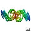
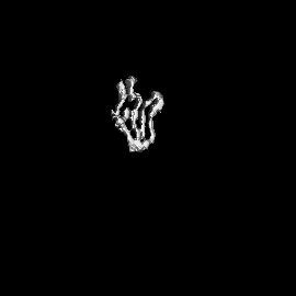
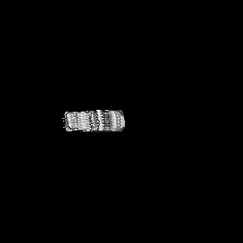
- EMDB-21201: Singlet Tau Fibril from Corticobasal Degeneration Human Brain Tissue -
+ Open data

- Basic information
| Entry | Database: EMDB / ID: EMD-21201 | |||||||||
|---|---|---|---|---|---|---|---|---|---|---|
| Title | Singlet Tau Fibril from Corticobasal Degeneration Human Brain Tissue | |||||||||
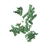
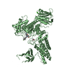
 Map data Map data | Singlet Tau Fibril from Corticobasal Degeneration Human Brain Tissue | |||||||||
 Sample Sample |
| |||||||||
 Keywords Keywords | Pathological amyloid fibril / cross-beta fold / parallel beta-sheets / PROTEIN FIBRIL | |||||||||
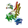
| Function / homology |  Function and homology information Function and homology informationplus-end-directed organelle transport along microtubule / histone-dependent DNA binding / negative regulation of protein localization to mitochondrion / neurofibrillary tangle / microtubule lateral binding / axonal transport / tubulin complex / positive regulation of protein localization to synapse / phosphatidylinositol bisphosphate binding / generation of neurons ...plus-end-directed organelle transport along microtubule / histone-dependent DNA binding / negative regulation of protein localization to mitochondrion / neurofibrillary tangle / microtubule lateral binding / axonal transport / tubulin complex / positive regulation of protein localization to synapse / phosphatidylinositol bisphosphate binding / generation of neurons / rRNA metabolic process / axonal transport of mitochondrion / axon development / regulation of mitochondrial fission / regulation of microtubule-based movement / central nervous system neuron development / intracellular distribution of mitochondria / regulation of chromosome organization / minor groove of adenine-thymine-rich DNA binding / lipoprotein particle binding / microtubule polymerization / negative regulation of mitochondrial membrane potential / regulation of microtubule polymerization / dynactin binding / apolipoprotein binding / protein polymerization / main axon / Caspase-mediated cleavage of cytoskeletal proteins / regulation of microtubule polymerization or depolymerization / negative regulation of mitochondrial fission / axolemma / glial cell projection / neurofibrillary tangle assembly / positive regulation of axon extension / regulation of cellular response to heat / positive regulation of microtubule polymerization / positive regulation of protein localization / Activation of AMPK downstream of NMDARs / positive regulation of superoxide anion generation / cytoplasmic microtubule organization / cellular response to brain-derived neurotrophic factor stimulus / regulation of calcium-mediated signaling / regulation of long-term synaptic depression / supramolecular fiber organization / axon cytoplasm / somatodendritic compartment / synapse assembly / nuclear periphery / phosphatidylinositol binding / astrocyte activation / protein phosphatase 2A binding / enzyme inhibitor activity / stress granule assembly / regulation of autophagy / regulation of microtubule cytoskeleton organization / cellular response to reactive oxygen species / microglial cell activation / cellular response to nerve growth factor stimulus / Hsp90 protein binding / protein homooligomerization / SH3 domain binding / PKR-mediated signaling / regulation of synaptic plasticity / synapse organization / microtubule cytoskeleton organization / response to lead ion / memory / neuron projection development / cytoplasmic ribonucleoprotein granule / cell-cell signaling / cellular response to heat / single-stranded DNA binding / microtubule cytoskeleton / growth cone / protein-folding chaperone binding / actin binding / double-stranded DNA binding / cell body / microtubule binding / sequence-specific DNA binding / amyloid fibril formation / dendritic spine / microtubule / learning or memory / protein-macromolecule adaptor activity / neuron projection / membrane raft / negative regulation of gene expression / axon / neuronal cell body / DNA damage response / dendrite / protein kinase binding / enzyme binding / mitochondrion / DNA binding / RNA binding / extracellular region / identical protein binding / nucleus Similarity search - Function | |||||||||
| Biological species |  Homo sapiens (human) Homo sapiens (human) | |||||||||
| Method | helical reconstruction / cryo EM / Resolution: 4.3 Å | |||||||||
 Authors Authors | Arakhamia T / Lee CE | |||||||||
| Funding support |  United States, 2 items United States, 2 items
| |||||||||
 Citation Citation | Journal: Cell / Year: 2020 Title: Posttranslational Modifications Mediate the Structural Diversity of Tauopathy Strains. Authors: Tamta Arakhamia / Christina E Lee / Yari Carlomagno / Duc M Duong / Sean R Kundinger / Kevin Wang / Dewight Williams / Michael DeTure / Dennis W Dickson / Casey N Cook / Nicholas T Seyfried ...Authors: Tamta Arakhamia / Christina E Lee / Yari Carlomagno / Duc M Duong / Sean R Kundinger / Kevin Wang / Dewight Williams / Michael DeTure / Dennis W Dickson / Casey N Cook / Nicholas T Seyfried / Leonard Petrucelli / Anthony W P Fitzpatrick / Abstract: Tau aggregation into insoluble filaments is the defining pathological hallmark of tauopathies. However, it is not known what controls the formation and templated seeding of strain-specific structures ...Tau aggregation into insoluble filaments is the defining pathological hallmark of tauopathies. However, it is not known what controls the formation and templated seeding of strain-specific structures associated with individual tauopathies. Here, we use cryo-electron microscopy (cryo-EM) to determine the structures of tau filaments from corticobasal degeneration (CBD) human brain tissue. Cryo-EM and mass spectrometry of tau filaments from CBD reveal that this conformer is heavily decorated with posttranslational modifications (PTMs), enabling us to map PTMs directly onto the structures. By comparing the structures and PTMs of tau filaments from CBD and Alzheimer's disease, it is found that ubiquitination of tau can mediate inter-protofilament interfaces. We propose a structure-based model in which cross-talk between PTMs influences tau filament structure, contributing to the structural diversity of tauopathy strains. Our approach establishes a framework for further elucidating the relationship between the structures of polymorphic fibrils, including their PTMs, and neurodegenerative disease. | |||||||||
| History |
|
- Structure visualization
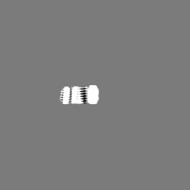
| Movie |
Movie viewer |
|---|---|
| Structure viewer | EM map: SurfViewMolmilJmol/JSmol |
| Supplemental images |

- Downloads & links
-EMDB archive
| Map data | emd_21201.map.gz | 510.4 KB | EMDB map data format | |
|---|---|---|---|---|
| Header (meta data) | emd-21201-v30.xmlemd-21201.xml | 11.9 KB 11.9 KB | Display Display | EMDB header |
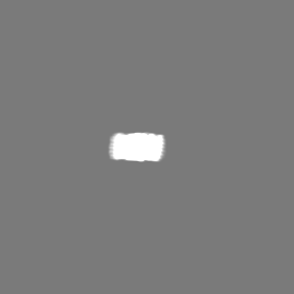
| Images | 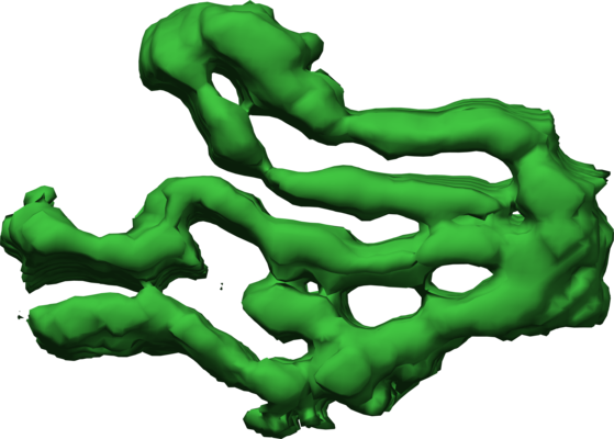 emd_21201.png emd_21201.png | 141.3 KB | ||
| Filedesc metadata | emd-21201.cif.gz | 5.3 KB | ||
| Archive directory |  http://ftp.pdbj.org/pub/emdb/structures/EMD-21201ftp://ftp.pdbj.org/pub/emdb/structures/EMD-21201 http://ftp.pdbj.org/pub/emdb/structures/EMD-21201ftp://ftp.pdbj.org/pub/emdb/structures/EMD-21201 | HTTPS FTP |
-Related structure data
| Related structure data | 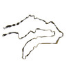 6vhaMC 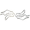 6vh7C 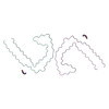 6vhlC 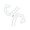 6vi3C M: atomic model generated by this map C: citing same article ( |
|---|---|
| Similar structure data |


-Links
| EMDB pages | EMDB (EBI/PDBe) / EMDataResource |
|---|---|
| Related items in Molecule of the Month |


-Map
| File | Download / File: emd_21201.map.gz / Format: CCP4 / Size: 75.1 MB / Type: IMAGE STORED AS FLOATING POINT NUMBER (4 BYTES) | ||||||||||||||||||||||||||||||||||||||||||||||||||||||||||||||||||||
|---|---|---|---|---|---|---|---|---|---|---|---|---|---|---|---|---|---|---|---|---|---|---|---|---|---|---|---|---|---|---|---|---|---|---|---|---|---|---|---|---|---|---|---|---|---|---|---|---|---|---|---|---|---|---|---|---|---|---|---|---|---|---|---|---|---|---|---|---|---|
| Annotation | Singlet Tau Fibril from Corticobasal Degeneration Human Brain Tissue | ||||||||||||||||||||||||||||||||||||||||||||||||||||||||||||||||||||
| Projections & slices | Image control
Images are generated by Spider. | ||||||||||||||||||||||||||||||||||||||||||||||||||||||||||||||||||||
| Voxel size | X=Y=Z: 1.414 Å | ||||||||||||||||||||||||||||||||||||||||||||||||||||||||||||||||||||
| Density |
| ||||||||||||||||||||||||||||||||||||||||||||||||||||||||||||||||||||
| Symmetry | Space group: 1 | ||||||||||||||||||||||||||||||||||||||||||||||||||||||||||||||||||||
| Details | EMDB XML:
CCP4 map header:
| ||||||||||||||||||||||||||||||||||||||||||||||||||||||||||||||||||||
 Z (Sec.)
 Y (Row.)
 X (Col.)


-Supplemental data
- Sample components
-Entire : Singlet Tau Fibril from Corticobasal Degeneration Human Brain Tissue
| Entire | Name: Singlet Tau Fibril from Corticobasal Degeneration Human Brain Tissue |
|---|---|
| Components |
|
-Supramolecule #1: Singlet Tau Fibril from Corticobasal Degeneration Human Brain Tissue
| Supramolecule | Name: Singlet Tau Fibril from Corticobasal Degeneration Human Brain Tissue type: tissue / ID: 1 / Parent: 0 / Macromolecule list: all |
|---|---|
| Source (natural) | Organism: Homo sapiens (human) |
-Macromolecule #1: Microtubule-associated protein tau
| Macromolecule | Name: Microtubule-associated protein tau / type: protein_or_peptide / ID: 1 / Number of copies: 3 / Enantiomer: LEVO |
|---|---|
| Source (natural) | Organism: Homo sapiens (human) |
| Molecular weight | Theoretical: 11.608375 KDa |
| Recombinant expression | Organism: Homo sapiens (human) |
| Sequence | String: KVQIINKKLD LSNVQSKCGS KDNIKHVPGG GSVQIVYKPV DLSKVTSKCG SLGNIHHKPG GGQVEVKSEK LDFKDRVQSK IGSLDNITH VPGGGNKKIE THKLTFRE UniProtKB: Microtubule-associated protein tau |
-Experimental details
-Structure determination
| Method | cryo EM |
|---|---|
 Processing Processing | helical reconstruction |
| Aggregation state | filament |
-Sample preparation
| Buffer | pH: 7.4 |
|---|---|
| Grid | Details: unspecified |
| Vitrification | Cryogen name: ETHANE |
- Electron microscopy
| Microscope | FEI TITAN KRIOS |
|---|---|
| Image recording | Film or detector model: GATAN K2 SUMMIT (4k x 4k) / Average electron dose: 60.0 e/Å2 |
| Electron beam | Acceleration voltage: 300 kV / Electron source:  FIELD EMISSION GUN FIELD EMISSION GUN |
| Electron optics | Illumination mode: FLOOD BEAM / Imaging mode: BRIGHT FIELD |
| Experimental equipment |  Model: Titan Krios / Image courtesy: FEI Company |
-Image processing
| Final reconstruction | Applied symmetry - Helical parameters - Δz: 4.8 Å Applied symmetry - Helical parameters - Δ&Phi: -0.85 ° Applied symmetry - Helical parameters - Axial symmetry: C1 (asymmetric) Resolution.type: BY AUTHOR / Resolution: 4.3 Å / Resolution method: FSC 0.143 CUT-OFF / Number images used: 9929 |
|---|---|
| CTF correction | Type: PHASE FLIPPING AND AMPLITUDE CORRECTION |
| Startup model | Type of model: NONE |
| Final angle assignment | Type: NOT APPLICABLE |
