 Movie
Movie Controller

[English] 日本語
 Yorodumi
- PDB-4osg: Klebsiella pneumoniae complexed with NADPH and 6-ethyl-5-[(3R)-3-... -
+ Open data

- Basic information
| Entry | Database: PDB / ID: 4osg | ||||||
|---|---|---|---|---|---|---|---|
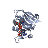
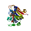
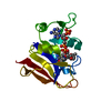
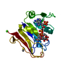
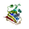
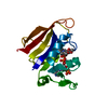
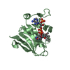
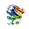
| Title | Klebsiella pneumoniae complexed with NADPH and 6-ethyl-5-[(3R)-3-[3-methoxyl-5-(pyridine-4-yl)phenyl]but-1-yn-1-yl]pyrimidine-2,4-diamine (UCP1006) | ||||||
 Components Components | Dihydrofolate reductase | ||||||
 Keywords Keywords | Oxidoreductase/Oxidoreductase inhibitor / Oxidoreductase / hydride shift / Oxidoreductase-Oxidoreductase inhibitor complex | ||||||
| Function / homology |  Function and homology information Function and homology informationDihydrofolate Reductase, subunit A / Dihydrofolate Reductase, subunit A / 3-Layer(aba) Sandwich / Alpha Beta Similarity search - Domain/homology | ||||||
| Biological species |  Klebsiella pneumoniae CG43 (bacteria) Klebsiella pneumoniae CG43 (bacteria) | ||||||
| Method |  X-RAY DIFFRACTION / SYNCHROTRON / MOLECULAR REPLACEMENT / Resolution: 2.7 Å X-RAY DIFFRACTION / SYNCHROTRON / MOLECULAR REPLACEMENT / Resolution: 2.7 Å | ||||||
 Authors Authors | Lamb, K.M. / Anderson, A.C. | ||||||
 Citation Citation | Journal: Antimicrob.Agents Chemother. / Year: 2014 Title: Crystal Structures of Klebsiella pneumoniae Dihydrofolate Reductase Bound to Propargyl-Linked Antifolates Reveal Features for Potency and Selectivity. Authors: Lamb, K.M. / Lombardo, M.N. / Alverson, J. / Priestley, N.D. / Wright, D.L. / Anderson, A.C. | ||||||
| History |
|
- Structure visualization
| Structure viewer | Molecule: MolmilJmol/JSmol |
|---|
- Downloads & links
-Download
| PDBx/mmCIF format | 4osg.cif.gz | 145.7 KB | Display | PDBx/mmCIF format |
|---|---|---|---|---|
| PDB format | pdb4osg.ent.gz | 116.9 KB | Display | PDB format |
| PDBx/mmJSON format | 4osg.json.gz | Tree view | PDBx/mmJSON format | |
| Others |  Other downloads Other downloads |
-Validation report
| Arichive directory | https://data.pdbj.org/pub/pdb/validation_reports/os/4osgftp://data.pdbj.org/pub/pdb/validation_reports/os/4osg | HTTPS FTP |
|---|
-Related structure data


-Links
 PDBj
PDBj


- Assembly
| Deposited unit | 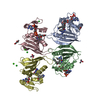
| ||||||||
|---|---|---|---|---|---|---|---|---|---|
| 1 | 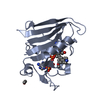
| ||||||||
| 2 | 
| ||||||||
| 3 | 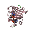
| ||||||||
| 4 | 
| ||||||||
| Unit cell |
|
-Components
-Protein , 1 types, 4 molecules ABCD
| #1: Protein | Mass: 18813.152 Da / Num. of mol.: 4 Source method: isolated from a genetically manipulated source Source: (gene. exp.) Klebsiella pneumoniae CG43 (bacteria) / Gene: folA, D364_00170 / Production host: |
|---|
-Non-polymers , 8 types, 31 molecules 


| #2: Chemical | ChemComp-NAP /  Mass: 743.405 Da / Num. of mol.: 4 / Source method: obtained synthetically / Formula: C21H28N7O17P3 Mass: 743.405 Da / Num. of mol.: 4 / Source method: obtained synthetically / Formula: C21H28N7O17P3#3: Chemical | ChemComp-06U /  Mass: 373.451 Da / Num. of mol.: 4 / Source method: obtained synthetically / Formula: C22H23N5O Mass: 373.451 Da / Num. of mol.: 4 / Source method: obtained synthetically / Formula: C22H23N5O#4: Chemical | ChemComp-EOH / |  Mass: 46.068 Da / Num. of mol.: 1 / Source method: obtained synthetically / Formula: C2H6O Mass: 46.068 Da / Num. of mol.: 1 / Source method: obtained synthetically / Formula: C2H6O#5: Chemical |  Mass: 136.989 Da / Num. of mol.: 2 / Source method: obtained synthetically / Formula: C2H6AsO2 Mass: 136.989 Da / Num. of mol.: 2 / Source method: obtained synthetically / Formula: C2H6AsO2#6: Chemical | ChemComp-CL /  Mass: 35.453 Da / Num. of mol.: 4 / Source method: obtained synthetically / Formula: Cl Mass: 35.453 Da / Num. of mol.: 4 / Source method: obtained synthetically / Formula: Cl#7: Chemical | ChemComp-PEG / |  Mass: 106.120 Da / Num. of mol.: 1 / Source method: obtained synthetically / Formula: C4H10O3 Mass: 106.120 Da / Num. of mol.: 1 / Source method: obtained synthetically / Formula: C4H10O3#8: Chemical |  Mass: 40.078 Da / Num. of mol.: 2 / Source method: obtained synthetically / Formula: Ca Mass: 40.078 Da / Num. of mol.: 2 / Source method: obtained synthetically / Formula: Ca#9: Water | ChemComp-HOH / | Mass: 18.015 Da / Num. of mol.: 13 / Source method: isolated from a natural source / Formula: H2O |
|---|
-Experimental details
-Experiment
| Experiment | Method: X-RAY DIFFRACTION / Number of used crystals: 1 |
|---|
- Sample preparation
| Crystal | Density Matthews: 2.62 Å3/Da / Density % sol: 53.02 % |
|---|---|
| Crystal grow | Temperature: 277 K / Method: vapor diffusion, sitting drop / pH: 7.4 Details: 100 mM cacodylate pH7.4, 125 mM sodium acetate, 25% (w/v) PEG 8000, 11 mM calcium chloride, 11 mM bentaine hydrochloride, VAPOR DIFFUSION, SITTING DROP, temperature 277K |
-Data collection
| Diffraction | Mean temperature: 100 K |
|---|---|
| Diffraction source | Source: SYNCHROTRON / Site: NSLS  / Beamline: X4A / Wavelength: 0.9789 Å / Beamline: X4A / Wavelength: 0.9789 Å |
| Detector | Type: ADSC QUANTUM 4 / Detector: CCD / Date: Nov 16, 2013 |
| Radiation | Monochromator: A KOHZU double crystal monochromator with a sagittally focused second crystal Protocol: SINGLE WAVELENGTH / Monochromatic (M) / Laue (L): M / Scattering type: x-ray |
| Radiation wavelength | Wavelength: 0.9789 Å / Relative weight: 1 |
| Reflection | Resolution: 2.7→39.27 Å / Num. all: 20988 / Num. obs: 19813 / % possible obs: 94.4 % / Observed criterion σ(F): 13.4 / Observed criterion σ(I): 13.4 / Redundancy: 1.67 % / Rmerge(I) obs: 0.049 / Rsym value: 0.07 / Net I/σ(I): 13.4 |
| Reflection shell | Resolution: 2.7→2.8 Å / Rmerge(I) obs: 0.331 / Mean I/σ(I) obs: 2.1 / Rsym value: 0.468 / % possible all: 95.1 |
- Processing
| Software |
| |||||||||||||||||||||||||||||||||||||||||||||||||||||||||||||||||||||||||||||||||||||||||||||||||||||||||
|---|---|---|---|---|---|---|---|---|---|---|---|---|---|---|---|---|---|---|---|---|---|---|---|---|---|---|---|---|---|---|---|---|---|---|---|---|---|---|---|---|---|---|---|---|---|---|---|---|---|---|---|---|---|---|---|---|---|---|---|---|---|---|---|---|---|---|---|---|---|---|---|---|---|---|---|---|---|---|---|---|---|---|---|---|---|---|---|---|---|---|---|---|---|---|---|---|---|---|---|---|---|---|---|---|---|---|
| Refinement | Method to determine structure: MOLECULAR REPLACEMENT / Resolution: 2.7→39.27 Å / σ(F): 2.7 / Phase error: 35.41 / Stereochemistry target values: TWIN_LSQ_F
| |||||||||||||||||||||||||||||||||||||||||||||||||||||||||||||||||||||||||||||||||||||||||||||||||||||||||
| Solvent computation | Shrinkage radii: 0.9 Å / VDW probe radii: 1.11 Å / Solvent model: FLAT BULK SOLVENT MODEL | |||||||||||||||||||||||||||||||||||||||||||||||||||||||||||||||||||||||||||||||||||||||||||||||||||||||||
| Refinement step | Cycle: LAST / Resolution: 2.7→39.27 Å
| |||||||||||||||||||||||||||||||||||||||||||||||||||||||||||||||||||||||||||||||||||||||||||||||||||||||||
| Refine LS restraints |
| |||||||||||||||||||||||||||||||||||||||||||||||||||||||||||||||||||||||||||||||||||||||||||||||||||||||||
| LS refinement shell |
|
