 Movie
Movie Controller

[English] 日本語
 Yorodumi
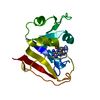
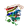
- PDB-1jom: THE CRYSTAL STRUCTURE OF THE BINARY COMPLEX BETWEEN FOLINIC ACID ... -
+ Open data

- Basic information
| Entry | Database: PDB / ID: 1jom | ||||||
|---|---|---|---|---|---|---|---|
| Title | THE CRYSTAL STRUCTURE OF THE BINARY COMPLEX BETWEEN FOLINIC ACID (LEUCOVORIN) AND E. COLI DIHYDROFOLATE REDUCTASE | ||||||
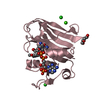
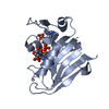
 Components Components | DIHYDROFOLATE REDUCTASE | ||||||
 Keywords Keywords | OXIDOREDUCTASE / METHOTREXATE RESISTANCE / ONE-CARBON METABOLISM OXIDOREDUCTASE | ||||||
| Function / homology |  Function and homology information Function and homology informationmethotrexate binding / dihydrofolic acid binding / 10-formyltetrahydrofolate biosynthetic process / response to methotrexate / folic acid biosynthetic process / folic acid binding / NADP+ binding / dihydrofolate metabolic process / dihydrofolate reductase / dihydrofolate reductase activity ...methotrexate binding / dihydrofolic acid binding / 10-formyltetrahydrofolate biosynthetic process / response to methotrexate / folic acid biosynthetic process / folic acid binding / NADP+ binding / dihydrofolate metabolic process / dihydrofolate reductase / dihydrofolate reductase activity / folic acid metabolic process / tetrahydrofolate biosynthetic process / NADPH binding / one-carbon metabolic process / NADP binding / response to xenobiotic stimulus / response to antibiotic / cytosol Similarity search - Function | ||||||
| Biological species |  | ||||||
| Method |  X-RAY DIFFRACTION / Resolution: 1.9 Å X-RAY DIFFRACTION / Resolution: 1.9 Å | ||||||
 Authors Authors | Reyes, V.M. / Lee, H. / Kraut, J. | ||||||
 Citation Citation | Journal: Biochemistry / Year: 1996 Title: Crystal structures of Escherichia coli dihydrofolate reductase complexed with 5-formyltetrahydrofolate (folinic acid) in two space groups: evidence for enolization of pteridine O4. Authors: Lee, H. / Reyes, V.M. / Kraut, J. #1: Journal: Biochemistry / Year: 1995Title: Isomorphous Crystal Structures of Escherichia Coli Dihydrofolate Reductase Complexed with Folate, 5-Deazafolate, and 5,10-Dideazatetrahydrofolate: Mechanistic Implications Authors: Reyes, V.M. / Sawaya, M.R. / Brown, K.A. / Kraut, J. #2: Journal: Biochemistry / Year: 1991Title: Crystal Structure of Unliganded Escherichia Coli Dihydrofolate Reductase. Ligand-Induced Conformational Changes and Cooperativity in Binding Authors: Bystroff, C. / Kraut, J. #3: Journal: Biochemistry / Year: 1990Title: Crystal Structures of Recombinant Human Dihydrofolate Reductase Complexed with Folate and 5-Deazafolate Authors: Davies II, J.F. / Delcamp, T.J. / Prendergast, N.J. / Ashford, V.A. / Freisheim, J.H. / Kraut, J. #4: Journal: Biochemistry / Year: 1990Title: Crystal Structures of Escherichia Coli Dihydrofolate Reductase: The Nadp+ Holoenzyme and the Folate.Nadp+ Ternary Complex. Substrate Binding and a Model for the Transition State Authors: Bystroff, C. / Oatley, S.J. / Kraut, J. #5: Journal: J.Biol.Chem. / Year: 1982Title: Crystal Structures of Escherichia Coli and Lactobacillus Casei Dihydrofolate Reductase Refined at 1.7 A Resolution. II. Environment of Bound Nadph and Implications for Catalysis Authors: Filman, D.J. / Bolin, J.T. / Matthews, D.A. / Kraut, J. #6: Journal: J.Biol.Chem. / Year: 1982Title: Crystal Structures of Escherichia Coli and Lactobacillus Casei Dihydrofolate Reductase Refined at 1.7 A Resolution. I. General Features and Binding of Methotrexate Authors: Bolin, J.T. / Filman, D.J. / Matthews, D.A. / Hamlin, R.C. / Kraut, J. | ||||||
| History |
|
- Structure visualization
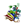
| Structure viewer | Molecule: MolmilJmol/JSmol |
|---|
- Downloads & links
-Download
| PDBx/mmCIF format | 1jom.cif.gz | 54.5 KB | Display | PDBx/mmCIF format |
|---|---|---|---|---|
| PDB format | pdb1jom.ent.gz | 38.8 KB | Display | PDB format |
| PDBx/mmJSON format | 1jom.json.gz | Tree view | PDBx/mmJSON format | |
| Others |  Other downloads Other downloads |
-Validation report
| Arichive directory | https://data.pdbj.org/pub/pdb/validation_reports/jo/1jomftp://data.pdbj.org/pub/pdb/validation_reports/jo/1jom | HTTPS FTP |
|---|
-Related structure data


-Links
 PDBj
PDBj


- Assembly
| Deposited unit | 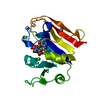
| ||||||||
|---|---|---|---|---|---|---|---|---|---|
| 1 |
| ||||||||
| Unit cell |
|
-Components
-Protein , 1 types, 1 molecules A
| #1: Protein | Mass: 18020.326 Da / Num. of mol.: 1 Source method: isolated from a genetically manipulated source Details: IN BINARY COMPLEX WITH FOLINIC ACID (LEUCOVORIN) / Source: (gene. exp.) |
|---|
-Non-polymers , 5 types, 217 molecules 


| #2: Chemical | ChemComp-CL /  Mass: 35.453 Da / Num. of mol.: 1 / Source method: obtained synthetically / Formula: Cl Mass: 35.453 Da / Num. of mol.: 1 / Source method: obtained synthetically / Formula: Cl | ||
|---|---|---|---|
| #3: Chemical | ChemComp-CA /  Mass: 40.078 Da / Num. of mol.: 1 / Source method: obtained synthetically / Formula: Ca Mass: 40.078 Da / Num. of mol.: 1 / Source method: obtained synthetically / Formula: Ca | ||
| #4: Chemical | ChemComp-FFO /  Mass: 473.439 Da / Num. of mol.: 1 / Source method: obtained synthetically / Formula: C20H23N7O7 Mass: 473.439 Da / Num. of mol.: 1 / Source method: obtained synthetically / Formula: C20H23N7O7 | ||
| #5: Chemical |  Mass: 46.068 Da / Num. of mol.: 3 / Source method: obtained synthetically / Formula: C2H6O Mass: 46.068 Da / Num. of mol.: 3 / Source method: obtained synthetically / Formula: C2H6O#6: Water | ChemComp-HOH / | Mass: 18.015 Da / Num. of mol.: 211 / Source method: isolated from a natural source / Formula: H2O |
-Experimental details
-Experiment
| Experiment | Method: X-RAY DIFFRACTION |
|---|
- Sample preparation
| Crystal | Density Matthews: 2.63 Å3/Da / Density % sol: 53.24 % | ||||||||||||||||||||||||||||||||||||
|---|---|---|---|---|---|---|---|---|---|---|---|---|---|---|---|---|---|---|---|---|---|---|---|---|---|---|---|---|---|---|---|---|---|---|---|---|---|
| Crystal grow | *PLUS Temperature: 4 ℃ / pH: 6.8 / Method: vapor diffusion | ||||||||||||||||||||||||||||||||||||
| Components of the solutions | *PLUS
|
-Data collection
| Radiation | Scattering type: x-ray |
|---|---|
| Radiation wavelength | Relative weight: 1 |
| Reflection | *PLUS Highest resolution: 1.9 Å / Num. obs: 15022 / % possible obs: 99.7 % / Redundancy: 7.7 % / Rmerge(I) obs: 0.068 |
- Processing
| Software | Name: TNT / Classification: refinement | ||||||||||||||||||||||||||||||
|---|---|---|---|---|---|---|---|---|---|---|---|---|---|---|---|---|---|---|---|---|---|---|---|---|---|---|---|---|---|---|---|
| Refinement | Highest resolution: 1.9 Å | ||||||||||||||||||||||||||||||
| Refinement step | Cycle: LAST / Highest resolution: 1.9 Å
| ||||||||||||||||||||||||||||||
| Refine LS restraints |
| ||||||||||||||||||||||||||||||
| Refinement | *PLUS Rfactor obs: 0.142 | ||||||||||||||||||||||||||||||
| Solvent computation | *PLUS | ||||||||||||||||||||||||||||||
| Displacement parameters | *PLUS |
