+ Open data
- Basic information
| Entry | Database: EMDB / ID: EMD-21170 | |||||||||
|---|---|---|---|---|---|---|---|---|---|---|
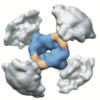
| Title | Tetrahedral nanoparticle T33_dn10 presenting BG505-SOSIP | |||||||||
 Map data Map data | BG505-SOSIP-T33_dn10-NP, Negative Stain EM map | |||||||||
 Sample Sample |
| |||||||||
| Biological species | synthetic construct (others) | |||||||||
| Method | single particle reconstruction / negative staining / Resolution: 25.67 Å | |||||||||
 Authors Authors | Ward AB / Antanasijevic A | |||||||||
| Funding support |  United States, 1 items United States, 1 items
| |||||||||
 Citation Citation |  Journal: Elife / Year: 2020 Journal: Elife / Year: 2020Title: Tailored design of protein nanoparticle scaffolds for multivalent presentation of viral glycoprotein antigens. Authors: George Ueda / Aleksandar Antanasijevic / Jorge A Fallas / William Sheffler / Jeffrey Copps / Daniel Ellis / Geoffrey B Hutchinson / Adam Moyer / Anila Yasmeen / Yaroslav Tsybovsky / Young- ...Authors: George Ueda / Aleksandar Antanasijevic / Jorge A Fallas / William Sheffler / Jeffrey Copps / Daniel Ellis / Geoffrey B Hutchinson / Adam Moyer / Anila Yasmeen / Yaroslav Tsybovsky / Young-Jun Park / Matthew J Bick / Banumathi Sankaran / Rebecca A Gillespie / Philip Jm Brouwer / Peter H Zwart / David Veesler / Masaru Kanekiyo / Barney S Graham / Rogier W Sanders / John P Moore / Per Johan Klasse / Andrew B Ward / Neil P King / David Baker /   Abstract: Multivalent presentation of viral glycoproteins can substantially increase the elicitation of antigen-specific antibodies. To enable a new generation of anti-viral vaccines, we designed self- ...Multivalent presentation of viral glycoproteins can substantially increase the elicitation of antigen-specific antibodies. To enable a new generation of anti-viral vaccines, we designed self-assembling protein nanoparticles with geometries tailored to present the ectodomains of influenza, HIV, and RSV viral glycoprotein trimers. We first designed trimers tailored for antigen fusion, featuring N-terminal helices positioned to match the C termini of the viral glycoproteins. Trimers that experimentally adopted their designed configurations were incorporated as components of tetrahedral, octahedral, and icosahedral nanoparticles, which were characterized by cryo-electron microscopy and assessed for their ability to present viral glycoproteins. Electron microscopy and antibody binding experiments demonstrated that the designed nanoparticles presented antigenically intact prefusion HIV-1 Env, influenza hemagglutinin, and RSV F trimers in the predicted geometries. This work demonstrates that antigen-displaying protein nanoparticles can be designed from scratch, and provides a systematic way to investigate the influence of antigen presentation geometry on the immune response to vaccination. | |||||||||
| History |
|
- Structure visualization
| Movie |
 Movie viewer Movie viewer |
|---|---|
| Structure viewer | EM map:  SurfView SurfView Molmil Molmil Jmol/JSmol Jmol/JSmol |
| Supplemental images |
- Downloads & links
-EMDB archive
| Map data |  emd_21170.map.gz emd_21170.map.gz | 71.1 MB |  EMDB map data format EMDB map data format | |
|---|---|---|---|---|
| Header (meta data) |  emd-21170-v30.xml emd-21170-v30.xml emd-21170.xml emd-21170.xml | 14.4 KB 14.4 KB | Display Display |  EMDB header EMDB header |
| Images |  emd_21170.png emd_21170.png | 144.4 KB | ||
| Archive directory |  http://ftp.pdbj.org/pub/emdb/structures/EMD-21170 http://ftp.pdbj.org/pub/emdb/structures/EMD-21170 ftp://ftp.pdbj.org/pub/emdb/structures/EMD-21170 ftp://ftp.pdbj.org/pub/emdb/structures/EMD-21170 | HTTPS FTP |
-Validation report
| Summary document |  emd_21170_validation.pdf.gz emd_21170_validation.pdf.gz | 77.2 KB | Display |  EMDB validaton report EMDB validaton report |
|---|---|---|---|---|
| Full document |  emd_21170_full_validation.pdf.gz emd_21170_full_validation.pdf.gz | 76.3 KB | Display | |
| Data in XML |  emd_21170_validation.xml.gz emd_21170_validation.xml.gz | 493 B | Display | |
| Arichive directory |  https://ftp.pdbj.org/pub/emdb/validation_reports/EMD-21170 https://ftp.pdbj.org/pub/emdb/validation_reports/EMD-21170 ftp://ftp.pdbj.org/pub/emdb/validation_reports/EMD-21170 ftp://ftp.pdbj.org/pub/emdb/validation_reports/EMD-21170 | HTTPS FTP |
-Related structure data
- Links
Links
| EMDB pages |  EMDB (EBI/PDBe) / EMDB (EBI/PDBe) /  EMDataResource EMDataResource |
|---|---|
| Related items in Molecule of the Month |
- Map
Map
| File |  Download / File: emd_21170.map.gz / Format: CCP4 / Size: 91.1 MB / Type: IMAGE STORED AS FLOATING POINT NUMBER (4 BYTES) Download / File: emd_21170.map.gz / Format: CCP4 / Size: 91.1 MB / Type: IMAGE STORED AS FLOATING POINT NUMBER (4 BYTES) | ||||||||||||||||||||||||||||||||||||||||||||||||||||||||||||||||||||
|---|---|---|---|---|---|---|---|---|---|---|---|---|---|---|---|---|---|---|---|---|---|---|---|---|---|---|---|---|---|---|---|---|---|---|---|---|---|---|---|---|---|---|---|---|---|---|---|---|---|---|---|---|---|---|---|---|---|---|---|---|---|---|---|---|---|---|---|---|---|
| Annotation | BG505-SOSIP-T33_dn10-NP, Negative Stain EM map | ||||||||||||||||||||||||||||||||||||||||||||||||||||||||||||||||||||
| Projections & slices | Image control
Images are generated by Spider. | ||||||||||||||||||||||||||||||||||||||||||||||||||||||||||||||||||||
| Voxel size | X=Y=Z: 2.05 Å | ||||||||||||||||||||||||||||||||||||||||||||||||||||||||||||||||||||
| Density |
| ||||||||||||||||||||||||||||||||||||||||||||||||||||||||||||||||||||
| Symmetry | Space group: 1 | ||||||||||||||||||||||||||||||||||||||||||||||||||||||||||||||||||||
| Details | EMDB XML:
CCP4 map header:
| ||||||||||||||||||||||||||||||||||||||||||||||||||||||||||||||||||||
-Supplemental data
- Sample components
-Entire : Tetrahedral nanoparticle, T33_dn10, presenting BG505-SOSIP
| Entire | Name: Tetrahedral nanoparticle, T33_dn10, presenting BG505-SOSIP |
|---|---|
| Components |
|
-Supramolecule #1: Tetrahedral nanoparticle, T33_dn10, presenting BG505-SOSIP
| Supramolecule | Name: Tetrahedral nanoparticle, T33_dn10, presenting BG505-SOSIP type: complex / ID: 1 / Parent: 0 / Macromolecule list: all / Details: 4 copies of BG505-SOSIP presented |
|---|---|
| Source (natural) | Organism: synthetic construct (others) |
| Recombinant expression | Organism:  Homo sapiens (human) / Recombinant strain: HEK293F / Recombinant plasmid: pPPI4 Homo sapiens (human) / Recombinant strain: HEK293F / Recombinant plasmid: pPPI4 |
-Macromolecule #1: BG505-SOSIP-T33_dn10A
| Macromolecule | Name: BG505-SOSIP-T33_dn10A / type: other / ID: 1 / Classification: other |
|---|---|
| Source (natural) | Organism: synthetic construct (others) |
| Sequence | String: MKRGLCCVLL LCGAVFVSPS QEIHARFRRG ARAENLWVTV YYGVPVWKDA ETTLFCASDA KAYETKKHNV WATHCCVPTD PNPQEIHLEN VTEEFNMWKN NMVEQMHTDI ISLWDQSLKP CVKLTPLCVT LQCTNVTNNI TDDMRGELKN CSFNMTTELR DKKQKVYSLF ...String: MKRGLCCVLL LCGAVFVSPS QEIHARFRRG ARAENLWVTV YYGVPVWKDA ETTLFCASDA KAYETKKHNV WATHCCVPTD PNPQEIHLEN VTEEFNMWKN NMVEQMHTDI ISLWDQSLKP CVKLTPLCVT LQCTNVTNNI TDDMRGELKN CSFNMTTELR DKKQKVYSLF YRLDVVQINE NQGNRSNNSN KEYRLINCNT SAITQACPKV SFEPIPIHYC APAGFAILKC KDKKFNGTGP CTNVSTVQCT HGIKPVVSTQ LLLNGSLAEE EVIIRSENIT NNAKNILVQL NESVQINCTR PNNNTVKSIR IGPGQWFYYT GDIIGDIRQA HCNVSKATWN ETLGKVVKQL RKHFGNNTII RFANSSGGDL EVTTHSFNCG GEFFYCNTSG LFNSTWISNT SVQGSNSTGS NDSITLPCRI KQIINMWQRI GQAMYAPPIQ GVIRCVSNIT GLILTRDGGS TNSTTETFRP GGGDMRDNWR SELYKYKVVK IEPLGVAPTR CKRRVVGRRR RRRAVGIGAV SLGFLGAAGS TMGAASMTLT VQARNLLSGI VQQQSNLLRA PECQQHLLKD THWGIKQLQA RVLAVEHYLR DQQLLGIWGC SGKLICCTNV PWNSSWSNRN LSEIWDNMTW LQWDKEISNY TQIIYGLLEE SQNQQEKNEQ GSGSGSGSGG EEAELAYLLG ELAYKLGEYR IAIRAYRIAL KRDPNNAEAW YNLGNAYYKQ GDYDEAIEYY QKALELDPNN AEAWYNLGNA YYKQGDYDEA IEYYEKALEL DPENLEALQN LLNAMDKQG |
| Recombinant expression | Organism:  Homo sapiens (human) Homo sapiens (human) |
-Macromolecule #2: T33_dn10B
| Macromolecule | Name: T33_dn10B / type: protein_or_peptide / ID: 2 / Enantiomer: LEVO |
|---|---|
| Source (natural) | Organism: synthetic construct (others) |
| Recombinant expression | Organism:  |
| Sequence | String: MIEEVVAEMI DILAESSKKS IEELARAADN KTTEKAVAEA IEEIARLATA AIQLIEALAK NLASEEFMAR AISAIAELAK KAIEAIYRLA DNHTTDTFMA RAIAAIANLA VTAILAIAAL ASNHTTEEFM ARAISAIAEL AKKAIEAIYR LADNHTTDKF MAAAIEAIAL ...String: MIEEVVAEMI DILAESSKKS IEELARAADN KTTEKAVAEA IEEIARLATA AIQLIEALAK NLASEEFMAR AISAIAELAK KAIEAIYRLA DNHTTDTFMA RAIAAIANLA VTAILAIAAL ASNHTTEEFM ARAISAIAEL AKKAIEAIYR LADNHTTDKF MAAAIEAIAL LATLAILAIA LLASNHTTEK FMARAIMAIA ILAAKAIEAI YRLADNHTSP TYIEKAIEAI EKIARKAIKA IEMLAKNITT EEYKEKAKKI IDIIRKLAKM AIKKLEDNRT LEHHHHHH |
-Experimental details
-Structure determination
| Method | negative staining |
|---|---|
 Processing Processing | single particle reconstruction |
| Aggregation state | particle |
- Sample preparation
| Concentration | 0.05 mg/mL | |||||||||
|---|---|---|---|---|---|---|---|---|---|---|
| Buffer | pH: 7.4 Component:
Details: TBS buffer, pH 7.4 | |||||||||
| Staining | Type: NEGATIVE / Material: Uranyl Formate Details: Sample diluted to 0.05 mg/mL. 3 uL was applied onto the grid, blotted off, and then stained with 2% uranyl formate for 60 seconds. | |||||||||
| Grid | Material: COPPER / Mesh: 400 / Support film - Material: CARBON / Support film - topology: CONTINUOUS | |||||||||
| Details | BG505-SOSIP-T33_dn10 nanoparticle was purified by SEC, diluted to 0.05mg/ml and loaded onto a grid. |
- Electron microscopy
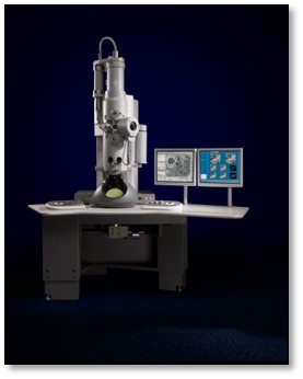
| Microscope | FEI TECNAI SPIRIT |
|---|---|
| Image recording | Film or detector model: TVIPS TEMCAM-F416 (4k x 4k) / Average electron dose: 25.0 e/Å2 |
| Electron beam | Acceleration voltage: 120 kV / Electron source: LAB6 |
| Electron optics | C2 aperture diameter: 100.0 µm / Illumination mode: FLOOD BEAM / Imaging mode: BRIGHT FIELD / Cs: 2.7 mm / Nominal defocus max: 1.5 µm / Nominal defocus min: 1.5 µm |
| Experimental equipment |  Model: Tecnai Spirit / Image courtesy: FEI Company |
 Movie
Movie Controller


 UCSF Chimera


 Z (Sec.)
 Y (Row.)
 X (Col.)


