 Movie
Movie Controller


+ Open data

- Basic information
| Entry | Database: EMDB / ID: EMD-20521 | |||||||||
|---|---|---|---|---|---|---|---|---|---|---|
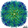
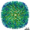
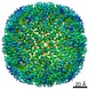
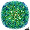
| Title | Horse spleen apoferritin light chain | |||||||||
 Map data Map data | Horse Spleen apoferritin LC sharpened map | |||||||||
 Sample Sample |
| |||||||||
 Keywords Keywords | Ferritin / apoferritin / octahedral / METAL BINDING PROTEIN | |||||||||
| Function / homology |  Function and homology information Function and homology informationferritin complex / autolysosome / ferric iron binding / autophagosome / iron ion transport / ferrous iron binding / cytoplasmic vesicle / intracellular iron ion homeostasis / iron ion binding / cytoplasm Similarity search - Function | |||||||||
| Biological species |  | |||||||||
| Method | single particle reconstruction / cryo EM / Resolution: 2.1 Å | |||||||||
 Authors Authors | Kopylov M / Kelley K | |||||||||
| Funding support |  United States, 1 items United States, 1 items
| |||||||||
 Citation Citation | Journal: To Be Published Title: Horse spleen apoferritin light chain structure at 2.1 Angstrom resolution Authors: Kopylov M / Kelley K / Yen LY / Rice WJ / Eng ET / Carragher B / Potter CS | |||||||||
| History |
|
- Structure visualization
| Movie |
Movie viewer |
|---|---|
| Structure viewer | EM map: SurfViewMolmilJmol/JSmol |
| Supplemental images |

- Downloads & links
-EMDB archive
| Map data | emd_20521.map.gz | 117.7 MB | EMDB map data format | |
|---|---|---|---|---|
| Header (meta data) | emd-20521-v30.xmlemd-20521.xml | 19.6 KB 19.6 KB | Display Display | EMDB header |
| FSC (resolution estimation) | emd_20521_fsc.xml | 13.1 KB | Display | FSC data file |
| Images | 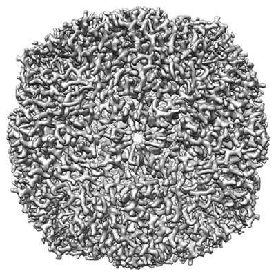 emd_20521.png emd_20521.png | 265.6 KB | ||
| Masks | emd_20521_msk_1.map | 125 MB | Mask map | |
| Filedesc metadata | emd-20521.cif.gz | 5.9 KB | ||
| Others | emd_20521_half_map_1.map.gzemd_20521_half_map_2.map.gz | 115.2 MB 115.2 MB | ||
| Archive directory |  http://ftp.pdbj.org/pub/emdb/structures/EMD-20521ftp://ftp.pdbj.org/pub/emdb/structures/EMD-20521 http://ftp.pdbj.org/pub/emdb/structures/EMD-20521ftp://ftp.pdbj.org/pub/emdb/structures/EMD-20521 | HTTPS FTP |
-Related structure data
| Related structure data |  6pxmMC M: atomic model generated by this map C: citing same article ( |
|---|---|
| Similar structure data |


-Links
| EMDB pages | EMDB (EBI/PDBe) / EMDataResource |
|---|---|
| Related items in Molecule of the Month |


-Map
| File | Download / File: emd_20521.map.gz / Format: CCP4 / Size: 125 MB / Type: IMAGE STORED AS FLOATING POINT NUMBER (4 BYTES) | ||||||||||||||||||||||||||||||||||||||||||||||||||||||||||||||||||||
|---|---|---|---|---|---|---|---|---|---|---|---|---|---|---|---|---|---|---|---|---|---|---|---|---|---|---|---|---|---|---|---|---|---|---|---|---|---|---|---|---|---|---|---|---|---|---|---|---|---|---|---|---|---|---|---|---|---|---|---|---|---|---|---|---|---|---|---|---|---|
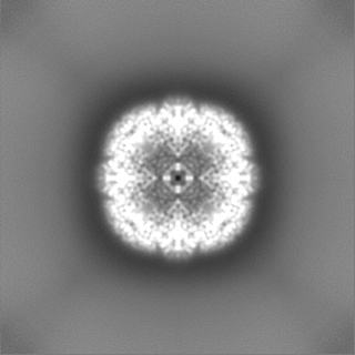
| Annotation | Horse Spleen apoferritin LC sharpened map | ||||||||||||||||||||||||||||||||||||||||||||||||||||||||||||||||||||
| Projections & slices | Image control
Images are generated by Spider. | ||||||||||||||||||||||||||||||||||||||||||||||||||||||||||||||||||||
| Voxel size | X=Y=Z: 0.832 Å | ||||||||||||||||||||||||||||||||||||||||||||||||||||||||||||||||||||
| Density |
| ||||||||||||||||||||||||||||||||||||||||||||||||||||||||||||||||||||
| Symmetry | Space group: 1 | ||||||||||||||||||||||||||||||||||||||||||||||||||||||||||||||||||||
| Details | EMDB XML:
CCP4 map header:
| ||||||||||||||||||||||||||||||||||||||||||||||||||||||||||||||||||||
 Z (Sec.)
 Y (Row.)
 X (Col.)


-Supplemental data
-Mask #1
| File | emd_20521_msk_1.map | ||||||||||||
|---|---|---|---|---|---|---|---|---|---|---|---|---|---|
| Projections & Slices |
| ||||||||||||
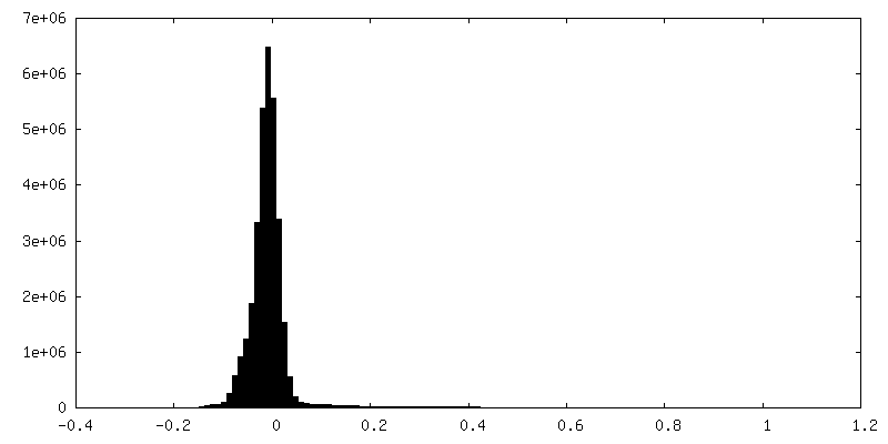
| Density Histograms |


-Half map: half A
| File | emd_20521_half_map_1.map | ||||||||||||
|---|---|---|---|---|---|---|---|---|---|---|---|---|---|
| Annotation | half A | ||||||||||||
| Projections & Slices |
| ||||||||||||
| Density Histograms |


-Half map: half B
| File | emd_20521_half_map_2.map | ||||||||||||
|---|---|---|---|---|---|---|---|---|---|---|---|---|---|
| Annotation | half B | ||||||||||||
| Projections & Slices |
| ||||||||||||
| Density Histograms |


- Sample components
-Entire : Horse spleen apoferritin light chain
| Entire | Name: Horse spleen apoferritin light chain |
|---|---|
| Components |
|
-Supramolecule #1: Horse spleen apoferritin light chain
| Supramolecule | Name: Horse spleen apoferritin light chain / type: complex / ID: 1 / Parent: 0 / Macromolecule list: all |
|---|---|
| Source (natural) | Organism: |
| Molecular weight | Theoretical: 480 KDa |
-Macromolecule #1: Ferritin light chain
| Macromolecule | Name: Ferritin light chain / type: protein_or_peptide / ID: 1 / Number of copies: 24 / Enantiomer: LEVO |
|---|---|
| Source (natural) | Organism: |
| Molecular weight | Theoretical: 20.003623 KDa |
| Sequence | String: MSSQIRQNYS TEVEAAVNRL VNLYLRASYT YLSLGFYFDR DDVALEGVCH FFRELAEEKR EGAERLLKMQ NQRGGRALFQ DLQKPSQDE WGTTLDAMKA AIVLEKSLNQ ALLDLHALGS AQADPHLCDF LESHFLDEEV KLIKKMGDHL TNIQRLVGSQ A GLGEYLFE RLTLKHD UniProtKB: Ferritin light chain |
-Experimental details
-Structure determination
| Method | cryo EM |
|---|---|
 Processing Processing | single particle reconstruction |
| Aggregation state | particle |
-Sample preparation
| Concentration | 7 mg/mL |
|---|---|
| Buffer | pH: 7.4 |
| Grid | Material: COPPER / Support film - Material: CARBON / Support film - topology: HOLEY / Pretreatment - Type: GLOW DISCHARGE / Pretreatment - Time: 20 sec. / Pretreatment - Atmosphere: AIR / Details: In-house-made nanowire grids |
| Vitrification | Cryogen name: ETHANE / Chamber humidity: 80 % / Chamber temperature: 298 K / Instrument: SPT LABTECH CHAMELEON / Details: Chameleon EP2 based on Spotiton. |
- Electron microscopy
| Microscope | FEI TITAN KRIOS |
|---|---|
| Image recording | Film or detector model: GATAN K2 SUMMIT (4k x 4k) / Detector mode: COUNTING / Number grids imaged: 1 / Number real images: 3885 / Average electron dose: 70.0 e/Å2 |
| Electron beam | Acceleration voltage: 300 kV / Electron source:  FIELD EMISSION GUN FIELD EMISSION GUN |
| Electron optics | C2 aperture diameter: 70.0 µm / Illumination mode: FLOOD BEAM / Imaging mode: BRIGHT FIELD / Cs: 2.7 mm / Nominal defocus max: -2.0 µm / Nominal defocus min: -1.0 µm / Nominal magnification: 29000 |
| Sample stage | Specimen holder model: FEI TITAN KRIOS AUTOGRID HOLDER / Cooling holder cryogen: NITROGEN |
| Experimental equipment |  Model: Titan Krios / Image courtesy: FEI Company |

