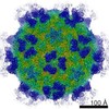
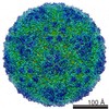
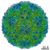
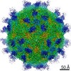
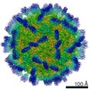
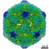
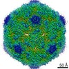
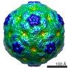
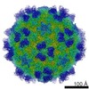
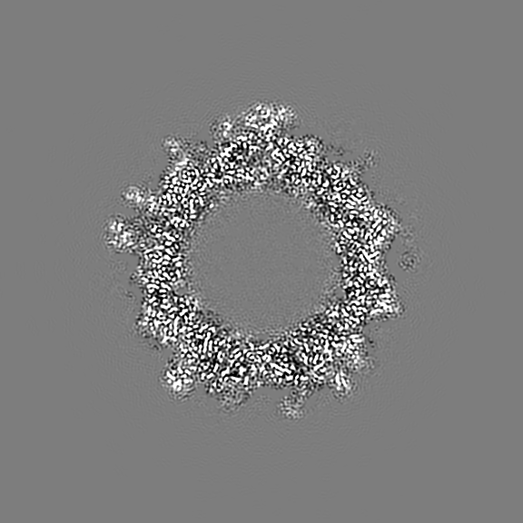
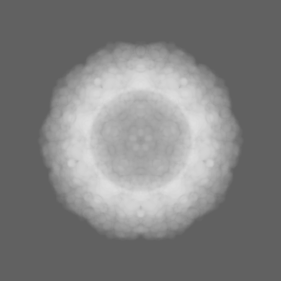
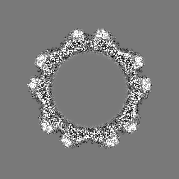
Journal: Proc Natl Acad Sci U S A / Year: 2020 Title: Cryo-EM structure of rhinovirus C15a bound to its cadherin-related protein 3 receptor. Authors: Yingyuan Sun / Kelly Watters / Marchel G Hill / Qianglin Fang / Yue Liu / Richard J Kuhn / Thomas Klose / Michael G Rossmann / Ann C Palmenberg / Abstract: Infection by (RV-C), a species of Picornaviridae , is strongly associated with childhood asthma exacerbations. Cellular binding and entry by all RV-C, which trigger these episodes, is mediated by ...Infection by (RV-C), a species of Picornaviridae , is strongly associated with childhood asthma exacerbations. Cellular binding and entry by all RV-C, which trigger these episodes, is mediated by the first extracellular domain (EC1) of cadherin-related protein 3 (CDHR3), a surface cadherin-like protein expressed primarily on the apical surfaces of ciliated airway epithelial cells. Although recombinant EC1 is a potent inhibitor of viral infection, there is no molecular description of this protein or its binding site on RV-C. Here we present cryo-electron microscopy (EM) data resolving the EC1 and EC1+2 domains of human CDHR3 complexed with viral isolate C15a. Structure-suggested residues contributing to required interfaces on both EC1 and C15a were probed and identified by mutagenesis studies with four different RV-C genotypes. In contrast to most other rhinoviruses, which bind intercellular adhesion molecule 1 receptors via a capsid protein VP1-specific fivefold canyon feature, the CDHR3 EC1 contacts C15a, and presumably all RV-Cs, in a unique cohesive footprint near the threefold vertex, encompassing residues primarily from viral protein VP3, but also from VP1 and VP2. The EC1+2 footprint on C15a is similar to that of EC1 alone but shows that steric hindrance imposed by EC2 would likely prevent multiprotein binding by the native receptor at any singular threefold vertex. Definition of the molecular interface between the RV-Cs and their receptors provides new avenues that can be explored for potential antiviral therapies.
History
Deposition
Jul 8, 2019
-
Header (metadata) release
Jul 24, 2019
-
Map release
Mar 11, 2020
-
Update
Mar 20, 2024
-
Current status
Mar 20, 2024
Processing site: RCSB / Status: Released
-
Structure visualization
Movie
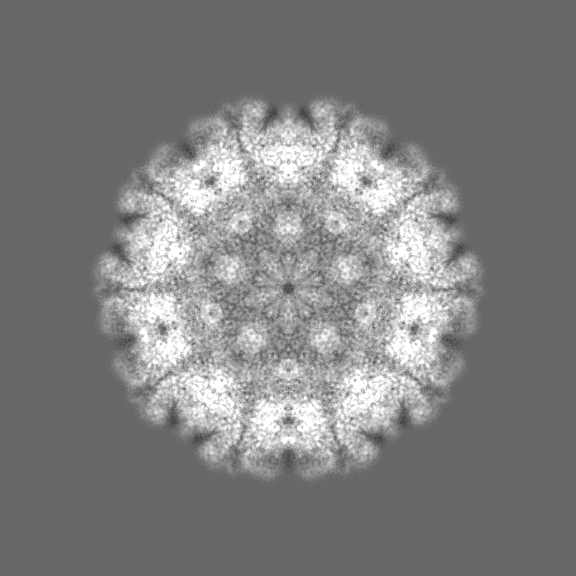
Surface view with section colored by density value
In the structure databanks used in Yorodumi, some data are registered as the other names, "COVID-19 virus" and "2019-nCoV". Here are the details of the virus and the list of structure data.
Jan 31, 2019. EMDB accession codes are about to change! (news from PDBe EMDB page)
EMDB accession codes are about to change! (news from PDBe EMDB page)
The allocation of 4 digits for EMDB accession codes will soon come to an end. Whilst these codes will remain in use, new EMDB accession codes will include an additional digit and will expand incrementally as the available range of codes is exhausted. The current 4-digit format prefixed with “EMD-” (i.e. EMD-XXXX) will advance to a 5-digit format (i.e. EMD-XXXXX), and so on. It is currently estimated that the 4-digit codes will be depleted around Spring 2019, at which point the 5-digit format will come into force.
The EM Navigator/Yorodumi systems omit the EMD- prefix.
Related info.:Q: What is EMD? / ID/Accession-code notation in Yorodumi/EM Navigator
Yorodumi is a browser for structure data from EMDB, PDB, SASBDB, etc.
This page is also the successor to EM Navigator detail page, and also detail information page/front-end page for Omokage search.
The word "yorodu" (or yorozu) is an old Japanese word meaning "ten thousand". "mi" (miru) is to see.
Related info.:EMDB / PDB / SASBDB / Comparison of 3 databanks / Yorodumi Search / Aug 31, 2016. New EM Navigator & Yorodumi / Yorodumi Papers / Jmol/JSmol / Function and homology information / Changes in new EM Navigator and Yorodumi
 Movie
Movie Controller


 Open data
 Basic information
 Map data
 Sample
 Keywords
 Function and homology information
 Homo sapiens (human) /
  Rhinovirus C
 Authors
 United States, 1 items
  Citation
 Structure visualization
 Downloads & links
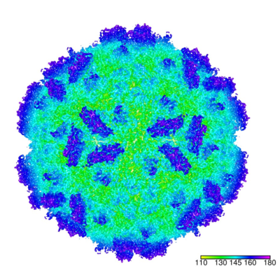 emd_20443.png
 http://ftp.pdbj.org/pub/emdb/structures/EMD-20443


 Z (Sec.)
 Y (Row.)
 X (Col.)


 Sample components
 Processing
 Electron microscopy
 FIELD EMISSION GUN


