 Movie
Movie Controller


+ Open data

- Basic information
| Entry | Database: PDB / ID: 5k0u | |||||||||
|---|---|---|---|---|---|---|---|---|---|---|
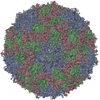
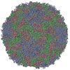
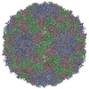
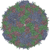
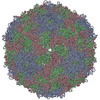
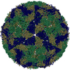
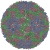
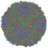
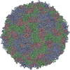
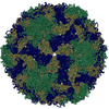
| Title | CryoEM structure of the full virion of a human rhinovirus C | |||||||||
 Components Components |
| |||||||||
 Keywords Keywords | VIRUS / jelly roll | |||||||||
| Function / homology |  Function and homology information Function and homology informationsymbiont-mediated suppression of host cytoplasmic pattern recognition receptor signaling pathway via inhibition of RIG-I activity / picornain 2A / symbiont-mediated suppression of host mRNA export from nucleus / symbiont genome entry into host cell via pore formation in plasma membrane / picornain 3C / T=pseudo3 icosahedral viral capsid / host cell cytoplasmic vesicle membrane / ribonucleoside triphosphate phosphatase activity / nucleoside-triphosphate phosphatase / channel activity ...symbiont-mediated suppression of host cytoplasmic pattern recognition receptor signaling pathway via inhibition of RIG-I activity / picornain 2A / symbiont-mediated suppression of host mRNA export from nucleus / symbiont genome entry into host cell via pore formation in plasma membrane / picornain 3C / T=pseudo3 icosahedral viral capsid / host cell cytoplasmic vesicle membrane / ribonucleoside triphosphate phosphatase activity / nucleoside-triphosphate phosphatase / channel activity / monoatomic ion transmembrane transport / DNA replication / RNA helicase activity / endocytosis involved in viral entry into host cell / symbiont-mediated activation of host autophagy / RNA-directed RNA polymerase / cysteine-type endopeptidase activity / viral RNA genome replication / RNA-directed RNA polymerase activity / virion attachment to host cell / host cell nucleus / structural molecule activity / DNA-templated transcription / proteolysis / RNA binding / zinc ion binding / ATP binding Similarity search - Function | |||||||||
| Biological species |  Rhinovirus C Rhinovirus C | |||||||||
| Method | ELECTRON MICROSCOPY / single particle reconstruction / cryo EM / Resolution: 2.79 Å | |||||||||
 Authors Authors | Liu, Y. / Hill, M.G. / Klose, T. / Chen, Z. / Watters, K.E. / Jiang, W. / Palmenberg, A.C. / Rossmann, M.G. | |||||||||
| Funding support |  United States, 2items United States, 2items
| |||||||||
 Citation Citation | Journal: Proc Natl Acad Sci U S A / Year: 2016 Title: Atomic structure of a rhinovirus C, a virus species linked to severe childhood asthma. Authors: Yue Liu / Marchel G Hill / Thomas Klose / Zhenguo Chen / Kelly Watters / Yury A Bochkov / Wen Jiang / Ann C Palmenberg / Michael G Rossmann / Abstract: Isolates of rhinovirus C (RV-C), a recently identified Enterovirus (EV) species, are the causative agents of severe respiratory infections among children and are linked to childhood asthma ...Isolates of rhinovirus C (RV-C), a recently identified Enterovirus (EV) species, are the causative agents of severe respiratory infections among children and are linked to childhood asthma exacerbations. The RV-C have been refractory to structure determination because they are difficult to propagate in vitro. Here, we report the cryo-EM atomic structures of the full virion and native empty particle (NEP) of RV-C15a. The virus has 60 "fingers" on the virus outer surface that probably function as dominant immunogens. Because the NEPs also display these fingers, they may have utility as vaccine candidates. A sequence-conserved surface depression adjacent to each finger forms a likely binding site for the sialic acid on its receptor. The RV-C, unlike other EVs, are resistant to capsid-binding antiviral compounds because the hydrophobic pocket in VP1 is filled with multiple bulky residues. These results define potential molecular determinants for designing antiviral therapeutics and vaccines. | |||||||||
| History |
|
- Structure visualization
| Movie |
Movie viewer |
|---|---|
| Structure viewer | Molecule: MolmilJmol/JSmol |
- Downloads & links
-Download
| PDBx/mmCIF format | 5k0u.cif.gz | 170.8 KB | Display | PDBx/mmCIF format |
|---|---|---|---|---|
| PDB format | pdb5k0u.ent.gz | 133.3 KB | Display | PDB format |
| PDBx/mmJSON format | 5k0u.json.gz | Tree view | PDBx/mmJSON format | |
| Others |  Other downloads Other downloads |
-Validation report
| Arichive directory | https://data.pdbj.org/pub/pdb/validation_reports/k0/5k0uftp://data.pdbj.org/pub/pdb/validation_reports/k0/5k0u | HTTPS FTP |
|---|
-Related structure data
| Related structure data | 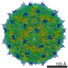 8189MC 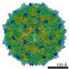 8184C 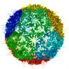 5jzgC M: map data used to model this data C: citing same article ( |
|---|---|
| Similar structure data |
-Links
 PDBj
PDBj

- Assembly
| Deposited unit | 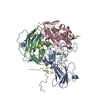
|
|---|---|
| 1 | x 60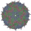
|
| 2 |
|
| 3 | x 5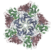
|
| 4 | x 6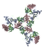
|
| 5 | 
|
| Symmetry | Point symmetry: (Schoenflies symbol: I (icosahedral)) |
-Components
| #1: Protein | Mass: 31802.623 Da / Num. of mol.: 1 / Fragment: UNP residues 568-846 / Mutation: T125K Source method: isolated from a genetically manipulated source Source: (gene. exp.) Rhinovirus C / Cell line: transduced HeLa expressing CDHR3 / Cell line (production host): HeLa WisL / Production host:  Homo sapiens (human) / References: UniProt: E5D8F2 Homo sapiens (human) / References: UniProt: E5D8F2 |
|---|---|
| #2: Protein | Mass: 25965.037 Da / Num. of mol.: 1 / Fragment: UNP residues 333-567 Source method: isolated from a genetically manipulated source Source: (gene. exp.) Rhinovirus C / Cell line: transduced HeLa expressing CDHR3 / Cell line (production host): HeLa WisL / Production host: Homo sapiens (human) / References: UniProt: E5D8F2 |
| #3: Protein | Mass: 29090.658 Da / Num. of mol.: 1 / Fragment: UNP residues 68-332 Source method: isolated from a genetically manipulated source Source: (gene. exp.) Rhinovirus C / Cell line: transduced HeLa expressing CDHR3 / Cell line (production host): HeLa WisL / Production host: Homo sapiens (human) / References: UniProt: E5D8F2 |
| #4: Protein | Mass: 7174.758 Da / Num. of mol.: 1 / Fragment: UNP residues 2-67 Source method: isolated from a genetically manipulated source Source: (gene. exp.) Rhinovirus C / Cell line: transduced HeLa expressing CDHR3 / Cell line (production host): HeLa WisL / Production host: Homo sapiens (human) / References: UniProt: E5D8F2 |
| #5: Water | ChemComp-HOH /  Mass: 18.015 Da / Num. of mol.: 60 / Source method: isolated from a natural source / Formula: H2O Mass: 18.015 Da / Num. of mol.: 60 / Source method: isolated from a natural source / Formula: H2O |
-Experimental details
-Experiment
| Experiment | Method: ELECTRON MICROSCOPY |
|---|---|
| EM experiment | Aggregation state: PARTICLE / 3D reconstruction method: single particle reconstruction |
- Sample preparation
| Component | Name: Rhinovirus C15a / Type: VIRUS Details: The cDNA of RV-C15 isolate was used to produce RNA transcripts in vitro, which were then used for transfection in HeLa WisL cells. The resultant recombinant RV-C15 virus was adapted in HeLa ...Details: The cDNA of RV-C15 isolate was used to produce RNA transcripts in vitro, which were then used for transfection in HeLa WisL cells. The resultant recombinant RV-C15 virus was adapted in HeLa cells expressing the RV-C receptor CDHR3 via multiple passage. The derivative was the RV-C15a virus. Entity ID: #1-#4 / Source: RECOMBINANT | ||||||||||||||||||||
|---|---|---|---|---|---|---|---|---|---|---|---|---|---|---|---|---|---|---|---|---|---|
| Molecular weight | Value: 8.3 MDa / Experimental value: NO | ||||||||||||||||||||
| Source (natural) | Organism: Rhinovirus C15a | ||||||||||||||||||||
| Source (recombinant) | Organism: Homo sapiens (human) / Cell: HeLa / Plasmid: pC15 | ||||||||||||||||||||
| Details of virus | Empty: NO / Enveloped: NO / Isolate: STRAIN / Type: VIRION | ||||||||||||||||||||
| Natural host | Organism: Homo sapiens | ||||||||||||||||||||
| Virus shell | Name: Capsid (p3) / Diameter: 300 nm / Triangulation number (T number): 3 | ||||||||||||||||||||
| Buffer solution | pH: 8 | ||||||||||||||||||||
| Buffer component |
| ||||||||||||||||||||
| Specimen | Conc.: 1 mg/ml / Embedding applied: NO / Shadowing applied: NO / Staining applied: NO / Vitrification applied: YES | ||||||||||||||||||||
| Specimen support | Grid material: COPPER / Grid mesh size: 400 divisions/in. / Grid type: Ultra thin Lacey carbon | ||||||||||||||||||||
| Vitrification | Instrument: GATAN CRYOPLUNGE 3 / Cryogen name: ETHANE / Humidity: 80 % / Chamber temperature: 298 K |
- Electron microscopy imaging
| Experimental equipment |  Model: Titan Krios / Image courtesy: FEI Company |
|---|---|
| Microscopy | Model: FEI TITAN KRIOS |
| Electron gun | Electron source:  FIELD EMISSION GUN / Accelerating voltage: 300 kV / Illumination mode: FLOOD BEAM FIELD EMISSION GUN / Accelerating voltage: 300 kV / Illumination mode: FLOOD BEAM |
| Electron lens | Mode: BRIGHT FIELD / Nominal magnification: 14000 X / Calibrated defocus min: 700 nm / Calibrated defocus max: 3500 nm / Cs: 2.7 mm / C2 aperture diameter: 100 µm / Alignment procedure: COMA FREE |
| Specimen holder | Cryogen: NITROGEN / Specimen holder model: FEI TITAN KRIOS AUTOGRID HOLDER |
| Image recording | Average exposure time: 14 sec. / Electron dose: 25.7 e/Å2 / Detector mode: SUPER-RESOLUTION / Film or detector model: GATAN K2 SUMMIT (4k x 4k) / Num. of grids imaged: 3 / Num. of real images: 8973 |
| Image scans | Movie frames/image: 70 / Used frames/image: 3-70 |
- Processing
| EM software |
| ||||||||||||||||||||||||||||||||||||||||||||||||||||||||||||
|---|---|---|---|---|---|---|---|---|---|---|---|---|---|---|---|---|---|---|---|---|---|---|---|---|---|---|---|---|---|---|---|---|---|---|---|---|---|---|---|---|---|---|---|---|---|---|---|---|---|---|---|---|---|---|---|---|---|---|---|---|---|
| CTF correction | Details: CTF correction was performed on the fly during 2D alignment and 3D reconstruction. Type: PHASE FLIPPING ONLY | ||||||||||||||||||||||||||||||||||||||||||||||||||||||||||||
| Particle selection | Num. of particles selected: 24882 Details: A mixed population of empty particles, full particles, and junk | ||||||||||||||||||||||||||||||||||||||||||||||||||||||||||||
| Symmetry | Point symmetry: I (icosahedral) | ||||||||||||||||||||||||||||||||||||||||||||||||||||||||||||
| 3D reconstruction | Resolution: 2.79 Å / Resolution method: FSC 0.143 CUT-OFF / Num. of particles: 8973 / Symmetry type: POINT | ||||||||||||||||||||||||||||||||||||||||||||||||||||||||||||
| Atomic model building | Protocol: RIGID BODY FIT / Space: REAL / Target criteria: Correlation coefficient |
