National Institutes of Health/National Institute of General Medical Sciences (NIH/NIGMS)
R01GM083118
United States
Citation
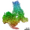
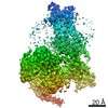
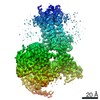
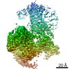
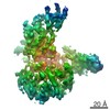
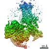
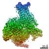
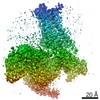
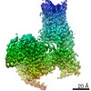
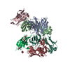
Journal: Science / Year: 2019 Title: Structures of the M1 and M2 muscarinic acetylcholine receptor/G-protein complexes. Authors: Shoji Maeda / Qianhui Qu / Michael J Robertson / Georgios Skiniotis / Brian K Kobilka / Abstract: Muscarinic acetylcholine receptors are G protein-coupled receptors that respond to acetylcholine and play important signaling roles in the nervous system. There are five muscarinic receptor subtypes ...Muscarinic acetylcholine receptors are G protein-coupled receptors that respond to acetylcholine and play important signaling roles in the nervous system. There are five muscarinic receptor subtypes (M1R to M5R), which, despite sharing a high degree of sequence identity in the transmembrane region, couple to different heterotrimeric GTP-binding proteins (G proteins) to transmit signals. M1R, M3R, and M5R couple to the G family, whereas M2R and M4R couple to the G family. Here, we present and compare the cryo-electron microscopy structures of M1R in complex with G and M2R in complex with G The M1R-G complex exhibits distinct features, including an extended transmembrane helix 5 and carboxyl-terminal receptor tail that interacts with G protein. Detailed analysis of these structures provides a framework for understanding the molecular determinants of G-protein coupling selectivity.
History
Deposition
Apr 9, 2019
-
Header (metadata) release
May 1, 2019
-
Map release
May 8, 2019
-
Update
Nov 20, 2024
-
Current status
Nov 20, 2024
Processing site: RCSB / Status: Released
-
Structure visualization
Movie
Surface view with section colored by density value
In the structure databanks used in Yorodumi, some data are registered as the other names, "COVID-19 virus" and "2019-nCoV". Here are the details of the virus and the list of structure data.
Jan 31, 2019. EMDB accession codes are about to change! (news from PDBe EMDB page)
EMDB accession codes are about to change! (news from PDBe EMDB page)
The allocation of 4 digits for EMDB accession codes will soon come to an end. Whilst these codes will remain in use, new EMDB accession codes will include an additional digit and will expand incrementally as the available range of codes is exhausted. The current 4-digit format prefixed with “EMD-” (i.e. EMD-XXXX) will advance to a 5-digit format (i.e. EMD-XXXXX), and so on. It is currently estimated that the 4-digit codes will be depleted around Spring 2019, at which point the 5-digit format will come into force.
The EM Navigator/Yorodumi systems omit the EMD- prefix.
Related info.:Q: What is EMD? / ID/Accession-code notation in Yorodumi/EM Navigator
Yorodumi is a browser for structure data from EMDB, PDB, SASBDB, etc.
This page is also the successor to EM Navigator detail page, and also detail information page/front-end page for Omokage search.
The word "yorodu" (or yorozu) is an old Japanese word meaning "ten thousand". "mi" (miru) is to see.
Related info.:EMDB / PDB / SASBDB / Comparison of 3 databanks / Yorodumi Search / Aug 31, 2016. New EM Navigator & Yorodumi / Yorodumi Papers / Jmol/JSmol / Function and homology information / Changes in new EM Navigator and Yorodumi
 Movie
Movie Controller


 Open data
 Basic information
 Map data
 Sample
 Keywords
 Function and homology information
 Homo sapiens (human) /
 Authors
 United States, 1 items
  Citation
 Structure visualization
 Downloads & links
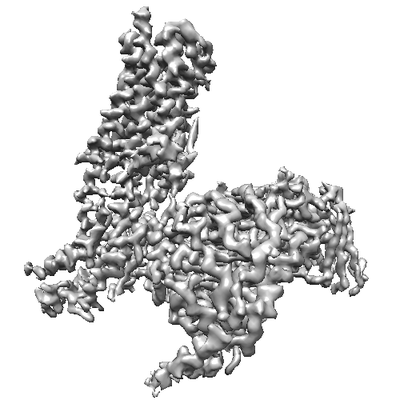 emd_20078.png
 http://ftp.pdbj.org/pub/emdb/structures/EMD-20078


 Z (Sec.)
 Y (Row.)
 X (Col.)


 Sample components
 Trichoplusia ni (cabbage looper)
 Spodoptera frugiperda (fall armyworm)

 Processing
 Electron microscopy
 FIELD EMISSION GUN
