 Movie
Movie Controller


+ Open data

- Basic information
| Entry | Database: PDB / ID: 6oij | ||||||
|---|---|---|---|---|---|---|---|
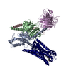
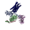
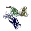
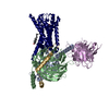
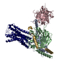
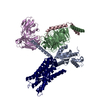
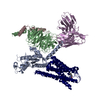
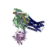
| Title | Muscarinic acetylcholine receptor 1-G11 protein complex | ||||||
 Components Components |
| ||||||
 Keywords Keywords | SIGNALING PROTEIN / G-protein coupled receptor-G-protein complex / neurotransmitter receptor | ||||||
| Function / homology |  Function and homology information Function and homology informationregulation of melanocyte differentiation / regulation of glial cell proliferation / saliva secretion / positive regulation of monoatomic ion transport / phospholipase C-activating G protein-coupled acetylcholine receptor signaling pathway / endothelin receptor signaling pathway / Muscarinic acetylcholine receptors / Fatty Acids bound to GPR40 (FFAR1) regulate insulin secretion / Acetylcholine regulates insulin secretion / G protein-coupled acetylcholine receptor activity ...regulation of melanocyte differentiation / regulation of glial cell proliferation / saliva secretion / positive regulation of monoatomic ion transport / phospholipase C-activating G protein-coupled acetylcholine receptor signaling pathway / endothelin receptor signaling pathway / Muscarinic acetylcholine receptors / Fatty Acids bound to GPR40 (FFAR1) regulate insulin secretion / Acetylcholine regulates insulin secretion / G protein-coupled acetylcholine receptor activity / developmental pigmentation / cholinergic synapse / phospholipase C-activating dopamine receptor signaling pathway / positive regulation of intracellular protein transport / cellular response to pH / PLC beta mediated events / phosphatidylinositol-4,5-bisphosphate phospholipase C activity / entrainment of circadian clock / cranial skeletal system development / neuromuscular synaptic transmission / adenylate cyclase-inhibiting G protein-coupled acetylcholine receptor signaling pathway / ligand-gated ion channel signaling pathway / regulation of locomotion / regulation of postsynaptic membrane potential / phototransduction, visible light / action potential / photoreceptor outer segment / G protein-coupled receptor signaling pathway, coupled to cyclic nucleotide second messenger / postsynaptic modulation of chemical synaptic transmission / enzyme regulator activity / Turbulent (oscillatory, disturbed) flow shear stress activates signaling by PIEZO1 and integrins in endothelial cells / axon terminus / skeletal system development / postsynaptic density membrane / G protein-coupled receptor binding / regulation of blood pressure / Schaffer collateral - CA1 synapse / cognition / G-protein beta/gamma-subunit complex binding / adenylate cyclase-modulating G protein-coupled receptor signaling pathway / positive regulation of insulin secretion / Olfactory Signaling Pathway / Activation of the phototransduction cascade / G protein-coupled acetylcholine receptor signaling pathway / G beta:gamma signalling through PLC beta / Presynaptic function of Kainate receptors / Thromboxane signalling through TP receptor / Activation of G protein gated Potassium channels / Inhibition of voltage gated Ca2+ channels via Gbeta/gamma subunits / G-protein activation / Glucagon signaling in metabolic regulation / Prostacyclin signalling through prostacyclin receptor / G beta:gamma signalling through CDC42 / Synthesis, secretion, and inactivation of Glucagon-like Peptide-1 (GLP-1) / G beta:gamma signalling through BTK / photoreceptor disc membrane / ADP signalling through P2Y purinoceptor 12 / Glucagon-type ligand receptors / Sensory perception of sweet, bitter, and umami (glutamate) taste / Adrenaline,noradrenaline inhibits insulin secretion / Vasopressin regulates renal water homeostasis via Aquaporins / Glucagon-like Peptide-1 (GLP1) regulates insulin secretion / G alpha (z) signalling events / cellular response to catecholamine stimulus / ADP signalling through P2Y purinoceptor 1 / ADORA2B mediated anti-inflammatory cytokines production / G beta:gamma signalling through PI3Kgamma / nervous system development / adenylate cyclase-activating dopamine receptor signaling pathway / Cooperation of PDCL (PhLP1) and TRiC/CCT in G-protein beta folding / GPER1 signaling / cellular response to prostaglandin E stimulus / heterotrimeric G-protein complex / G alpha (12/13) signalling events / Inactivation, recovery and regulation of the phototransduction cascade / G-protein beta-subunit binding / extracellular vesicle / sensory perception of taste / heart development / Thrombin signalling through proteinase activated receptors (PARs) / signaling receptor complex adaptor activity / retina development in camera-type eye / GTPase binding / fibroblast proliferation / G protein activity / presynaptic membrane / Ca2+ pathway / High laminar flow shear stress activates signaling by PIEZO1 and PECAM1:CDH5:KDR in endothelial cells / G alpha (i) signalling events / G alpha (s) signalling events / phospholipase C-activating G protein-coupled receptor signaling pathway / G alpha (q) signalling events / Hydrolases; Acting on acid anhydrides; Acting on GTP to facilitate cellular and subcellular movement / chemical synaptic transmission / Ras protein signal transduction / Extra-nuclear estrogen signaling / cell population proliferation / G protein-coupled receptor signaling pathway / lysosomal membrane / GTPase activity Similarity search - Function | ||||||
| Biological species |  Homo sapiens (human) Homo sapiens (human) | ||||||
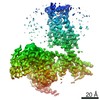
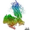
| Method | ELECTRON MICROSCOPY / single particle reconstruction / cryo EM / Resolution: 3.3 Å | ||||||
 Authors Authors | Maeda, S. / Qianhui, Q. / Skiniotis, G. / Kobilka, B. | ||||||
| Funding support |  United States, 1items United States, 1items
| ||||||
 Citation Citation | Journal: Science / Year: 2019 Title: Structures of the M1 and M2 muscarinic acetylcholine receptor/G-protein complexes. Authors: Shoji Maeda / Qianhui Qu / Michael J Robertson / Georgios Skiniotis / Brian K Kobilka / Abstract: Muscarinic acetylcholine receptors are G protein-coupled receptors that respond to acetylcholine and play important signaling roles in the nervous system. There are five muscarinic receptor subtypes ...Muscarinic acetylcholine receptors are G protein-coupled receptors that respond to acetylcholine and play important signaling roles in the nervous system. There are five muscarinic receptor subtypes (M1R to M5R), which, despite sharing a high degree of sequence identity in the transmembrane region, couple to different heterotrimeric GTP-binding proteins (G proteins) to transmit signals. M1R, M3R, and M5R couple to the G family, whereas M2R and M4R couple to the G family. Here, we present and compare the cryo-electron microscopy structures of M1R in complex with G and M2R in complex with G The M1R-G complex exhibits distinct features, including an extended transmembrane helix 5 and carboxyl-terminal receptor tail that interacts with G protein. Detailed analysis of these structures provides a framework for understanding the molecular determinants of G-protein coupling selectivity. | ||||||
| History |
|
- Structure visualization
| Movie |
Movie viewer |
|---|---|
| Structure viewer | Molecule: MolmilJmol/JSmol |
- Downloads & links
-Download
| PDBx/mmCIF format | 6oij.cif.gz | 220.1 KB | Display | PDBx/mmCIF format |
|---|---|---|---|---|
| PDB format | pdb6oij.ent.gz | 166.5 KB | Display | PDB format |
| PDBx/mmJSON format | 6oij.json.gz | Tree view | PDBx/mmJSON format | |
| Others |  Other downloads Other downloads |
-Validation report
| Arichive directory | https://data.pdbj.org/pub/pdb/validation_reports/oi/6oijftp://data.pdbj.org/pub/pdb/validation_reports/oi/6oij | HTTPS FTP |
|---|
-Related structure data
| Related structure data |  20078MC 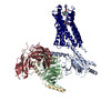 6oikC M: map data used to model this data C: citing same article ( |
|---|---|
| Similar structure data |

-Links
 PDBj
PDBj


- Assembly
| Deposited unit | 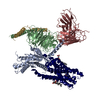
|
|---|---|
| 1 |
|
-Components
-Guanine nucleotide-binding protein ... , 3 types, 3 molecules ABG
| #1: Protein | Mass: 41271.086 Da / Num. of mol.: 1 Mutation: chimeric protein between G-alpha1i(1-29) and G-alpha11(30-) Source method: isolated from a genetically manipulated source Source: (gene. exp.) Homo sapiens (human) / Gene: GNAI1, GNA11, GA11 / Production host:  Trichoplusia ni (cabbage looper) / References: UniProt: A0A3B3ITX3, UniProt: P29992 Trichoplusia ni (cabbage looper) / References: UniProt: A0A3B3ITX3, UniProt: P29992 |
|---|---|
| #4: Protein | Mass: 37728.152 Da / Num. of mol.: 1 Source method: isolated from a genetically manipulated source Source: (gene. exp.) Homo sapiens (human) / Gene: GNB1 / Production host: Trichoplusia ni (cabbage looper) / References: UniProt: P62873 |
| #5: Protein | Mass: 7861.143 Da / Num. of mol.: 1 Source method: isolated from a genetically manipulated source Source: (gene. exp.) Homo sapiens (human) / Gene: GNG2 / Production host: Trichoplusia ni (cabbage looper) / References: UniProt: P59768 |
-Protein / Antibody , 2 types, 2 molecules RH
| #2: Protein | Mass: 41241.262 Da / Num. of mol.: 1 / Mutation: N11Q, N21Q Source method: isolated from a genetically manipulated source Source: (gene. exp.) Homo sapiens (human) / Gene: CHRM1 / Production host:  Spodoptera frugiperda (fall armyworm) / References: UniProt: P11229 Spodoptera frugiperda (fall armyworm) / References: UniProt: P11229 |
|---|---|
| #3: Antibody | Mass: 27340.482 Da / Num. of mol.: 1 Source method: isolated from a genetically manipulated source Source: (gene. exp.) Trichoplusia ni (cabbage looper) |
-Non-polymers , 2 types, 3 molecules 

| #6: Chemical | ChemComp-IXO /  Mass: 197.254 Da / Num. of mol.: 1 / Source method: obtained synthetically / Formula: C10H17N2O2 Mass: 197.254 Da / Num. of mol.: 1 / Source method: obtained synthetically / Formula: C10H17N2O2 |
|---|---|
| #7: Chemical |  Mass: 486.726 Da / Num. of mol.: 2 / Source method: obtained synthetically / Formula: C31H50O4 Mass: 486.726 Da / Num. of mol.: 2 / Source method: obtained synthetically / Formula: C31H50O4 |
-Details
| Has protein modification | Y |
|---|
-Experimental details
-Experiment
| Experiment | Method: ELECTRON MICROSCOPY |
|---|---|
| EM experiment | Aggregation state: PARTICLE / 3D reconstruction method: single particle reconstruction |
- Sample preparation
| Component |
| ||||||||||||||||||||||||
|---|---|---|---|---|---|---|---|---|---|---|---|---|---|---|---|---|---|---|---|---|---|---|---|---|---|
| Molecular weight | Value: 0.15 MDa / Experimental value: NO | ||||||||||||||||||||||||
| Source (natural) |
| ||||||||||||||||||||||||
| Source (recombinant) |
| ||||||||||||||||||||||||
| Buffer solution | pH: 7.5 | ||||||||||||||||||||||||
| Specimen | Embedding applied: NO / Shadowing applied: NO / Staining applied: NO / Vitrification applied: YES | ||||||||||||||||||||||||
| Specimen support | Details: unspecified | ||||||||||||||||||||||||
| Vitrification | Cryogen name: ETHANE |
- Electron microscopy imaging
| Experimental equipment |  Model: Titan Krios / Image courtesy: FEI Company |
|---|---|
| Microscopy | Model: FEI TITAN KRIOS |
| Electron gun | Electron source:  FIELD EMISSION GUN / Accelerating voltage: 300 kV / Illumination mode: FLOOD BEAM FIELD EMISSION GUN / Accelerating voltage: 300 kV / Illumination mode: FLOOD BEAM |
| Electron lens | Mode: BRIGHT FIELD |
| Image recording | Electron dose: 7 e/Å2 / Detector mode: COUNTING / Film or detector model: GATAN K2 SUMMIT (4k x 4k) |
- Processing
| EM software | Name: RELION / Version: 2.1 / Category: image acquisition |
|---|---|
| CTF correction | Type: PHASE FLIPPING AND AMPLITUDE CORRECTION |
| Symmetry | Point symmetry: C1 (asymmetric) |
| 3D reconstruction | Resolution: 3.3 Å / Resolution method: FSC 0.143 CUT-OFF / Num. of particles: 277988 / Symmetry type: POINT |
