 ムービー
ムービー コントローラー


+ データを開く

- 基本情報
基本情報
| 登録情報 | データベース: EMDB / ID: EMD-1995 | |||||||||
|---|---|---|---|---|---|---|---|---|---|---|
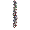
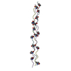
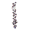
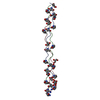
| タイトル | Electron density map of a composite coiled-coil fibril comprising multiple self-assembling fibre peptides. | |||||||||
 マップデータ マップデータ | This is a map of a coiled coil derived from a micrograph of a 3D protein fibre processed using 2dx | |||||||||
 試料 試料 |
| |||||||||
 キーワード キーワード | CryoTEM / coiled coil / fibrous proteins / protein design / self-assembly | |||||||||
| 生物種 | synthetic construct (人工物) | |||||||||
| 手法 | 電子線結晶学 / クライオ電子顕微鏡法 / 解像度: 8.0 Å | |||||||||
 データ登録者 データ登録者 | Sharp TH / Bruning M / Mantell J / Sessions RB / Thomson AR / Zaccai NR / Brady RL / Verkade P / Woolfson DN | |||||||||
 引用 引用 | ジャーナル: Proc Natl Acad Sci U S A / 年: 2012 タイトル: Cryo-transmission electron microscopy structure of a gigadalton peptide fiber of de novo design. 著者: Thomas H Sharp / Marc Bruning / Judith Mantell / Richard B Sessions / Andrew R Thomson / Nathan R Zaccai / R Leo Brady / Paul Verkade / Derek N Woolfson /  要旨: Nature presents various protein fibers that bridge the nanometer to micrometer regimes. These structures provide inspiration for the de novo design of biomimetic assemblies, both to address ...Nature presents various protein fibers that bridge the nanometer to micrometer regimes. These structures provide inspiration for the de novo design of biomimetic assemblies, both to address difficulties in studying and understanding natural systems, and to provide routes to new biomaterials with potential applications in nanotechnology and medicine. We have designed a self-assembling fiber system, the SAFs, in which two small α-helical peptides are programmed to form a dimeric coiled coil and assemble in a controlled manner. The resulting fibers are tens of nm wide and tens of μm long, and, therefore, comprise millions of peptides to give gigadalton supramolecular structures. Here, we describe the structure of the SAFs determined to approximately 8 Å resolution using cryotransmission electron microscopy. Individual micrographs show clear ultrastructure that allowed direct interpretation of the packing of individual α-helices within the fibers, and the construction of a 3D electron density map. Furthermore, a model was derived using the cryotransmission electron microscopy data and side chains taken from a 2.3 Å X-ray crystal structure of a peptide building block incapable of forming fibers. This was validated using single-particle analysis techniques, and was stable in prolonged molecular-dynamics simulation, confirming its structural viability. The level of self-assembly and self-organization in the SAFs is unprecedented for a designed peptide-based material, particularly for a system of considerably reduced complexity compared with natural proteins. This structural insight is a unique high-resolution description of how α-helical fibrils pack into larger protein fibers, and provides a basis for the design and engineering of future biomaterials. | |||||||||
| 履歴 |
|
- 構造の表示
構造の表示
| ムービー |
 ムービービューア ムービービューア |
|---|---|
| 構造ビューア | EMマップ: SurfViewMolmilJmol/JSmol |
| 添付画像 |
 UCSF Chimera
- ダウンロードとリンク
-EMDBアーカイブ
| マップデータ | emd_1995.map.gz | 3.6 MB | EMDBマップデータ形式 | |
|---|---|---|---|---|
| ヘッダ (付随情報) | emd-1995-v30.xmlemd-1995.xml | 11.5 KB 11.5 KB | 表示 表示 | EMDBヘッダ |
| 画像 | 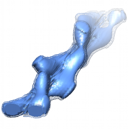 SAF.png SAF.png | 243.3 KB | ||
| アーカイブディレクトリ |  http://ftp.pdbj.org/pub/emdb/structures/EMD-1995ftp://ftp.pdbj.org/pub/emdb/structures/EMD-1995 http://ftp.pdbj.org/pub/emdb/structures/EMD-1995ftp://ftp.pdbj.org/pub/emdb/structures/EMD-1995 | HTTPS FTP |
-検証レポート
| 文書・要旨 | emd_1995_validation.pdf.gz | 183.2 KB | 表示 | EMDB検証レポート |
|---|---|---|---|---|
| 文書・詳細版 | emd_1995_full_validation.pdf.gz | 182.4 KB | 表示 | |
| XML形式データ | emd_1995_validation.xml.gz | 4.7 KB | 表示 | |
| アーカイブディレクトリ | https://ftp.pdbj.org/pub/emdb/validation_reports/EMD-1995ftp://ftp.pdbj.org/pub/emdb/validation_reports/EMD-1995 | HTTPS FTP |
-関連構造データ

-リンク
| EMDBのページ | EMDB (EBI/PDBe) / EMDataResource |
|---|
-マップ
| ファイル | ダウンロード / ファイル: emd_1995.map.gz / 形式: CCP4 / 大きさ: 4.8 MB / タイプ: IMAGE STORED AS FLOATING POINT NUMBER (4 BYTES) | ||||||||||||||||||||||||||||||||||||||||||||||||||||||||||||||||||||
|---|---|---|---|---|---|---|---|---|---|---|---|---|---|---|---|---|---|---|---|---|---|---|---|---|---|---|---|---|---|---|---|---|---|---|---|---|---|---|---|---|---|---|---|---|---|---|---|---|---|---|---|---|---|---|---|---|---|---|---|---|---|---|---|---|---|---|---|---|---|
| 注釈 | This is a map of a coiled coil derived from a micrograph of a 3D protein fibre processed using 2dx | ||||||||||||||||||||||||||||||||||||||||||||||||||||||||||||||||||||
| 投影像・断面図 | 画像のコントロール
画像は Spider により作成 これらの図は立方格子座標系で作成されたものです | ||||||||||||||||||||||||||||||||||||||||||||||||||||||||||||||||||||
| ボクセルのサイズ | X=Y=Z: 0.627 Å | ||||||||||||||||||||||||||||||||||||||||||||||||||||||||||||||||||||
| 密度 |
| ||||||||||||||||||||||||||||||||||||||||||||||||||||||||||||||||||||
| 対称性 | 空間群: 1 | ||||||||||||||||||||||||||||||||||||||||||||||||||||||||||||||||||||
| 詳細 | EMDB XML:
CCP4マップ ヘッダ情報:
| ||||||||||||||||||||||||||||||||||||||||||||||||||||||||||||||||||||
 Z (Sec.)
 Y (Row.)
 X (Col.)


-添付データ
- 試料の構成要素
-全体 : Self-assembling peptide fibre
| 全体 | 名称: Self-assembling peptide fibre |
|---|---|
| 要素 |
|
-超分子 #1000: Self-assembling peptide fibre
| 超分子 | 名称: Self-assembling peptide fibre / タイプ: sample / ID: 1000 / 詳細: Samples contains approximately 30,000,000 peptides 集合状態: Many millions of peptides self-assemble to form a gigadalton peptide fibre Number unique components: 2 |
|---|
-分子 #1: KIAALKQKIASLKQEIDALEYENDALEQ
| 分子 | 名称: KIAALKQKIASLKQEIDALEYENDALEQ / タイプ: protein_or_peptide / ID: 1 / Name.synonym: Self-assembling fibre 詳細: Peptides were synthesized by microwave-assisted solid-phase peptide synthesis using standard HBTU activation. They heterodimerize to form offset dimeric coiled coils with complementary sticky ...詳細: Peptides were synthesized by microwave-assisted solid-phase peptide synthesis using standard HBTU activation. They heterodimerize to form offset dimeric coiled coils with complementary sticky ends that assemble to form extended coiled coil fibrils. These fibrils pack laterally to generate large proteinaceous fibres on average 82 nm in diameter and 42 micrometers in length. 組換発現: No / データベース: NCBI |
|---|---|
| 由来(天然) | 生物種: synthetic construct (人工物) |
| 分子量 | 理論値: 3.17 KDa |
-分子 #2: KIRRLKQKNARLKQEIAALEYEIAALEQ
| 分子 | 名称: KIRRLKQKNARLKQEIAALEYEIAALEQ / タイプ: protein_or_peptide / ID: 2 / Name.synonym: Self-assembling fibre 詳細: Peptides were synthesized by microwave-assisted solid-phase peptide synthesis using standard HBTU activation. They heterodimerize to form offset dimeric coiled coils with complementary sticky ...詳細: Peptides were synthesized by microwave-assisted solid-phase peptide synthesis using standard HBTU activation. They heterodimerize to form offset dimeric coiled coils with complementary sticky ends that assemble to form extended coiled coil fibrils. These fibrils pack laterally to generate large proteinaceous fibres on average 82 nm in diameter and 42 micrometers in length. 組換発現: No / データベース: NCBI |
|---|---|
| 由来(天然) | 生物種: synthetic construct (人工物) |
| 分子量 | 理論値: 3.32 KDa |
-実験情報
-構造解析
| 手法 | クライオ電子顕微鏡法 |
|---|---|
 解析 解析 | 電子線結晶学 |
| 試料の集合状態 | 2D array |
-試料調製
| 濃度 | 0.325 mg/mL |
|---|---|
| 緩衝液 | pH: 7.4 / 詳細: 10 mM MOPS |
| グリッド | 詳細: Lacey-carbon grid |
| 凍結 | 凍結剤: ETHANE / チャンバー内湿度: 100 % / チャンバー内温度: 98 K / 装置: OTHER / 詳細: Vitrification instrument: Vitrobot / 手法: Blot 1 sec. |
- 電子顕微鏡法
| 顕微鏡 | FEI TECNAI 20 |
|---|---|
| 詳細 | Low dose software used |
| 日付 | 2010年4月20日 |
| 撮影 | デジタル化 - サンプリング間隔: 11 µm / 実像数: 1 / 平均電子線量: 10 e/Å2 |
| Tilt angle min | 0 |
| Tilt angle max | 0 |
| 電子線 | 加速電圧: 200 kV / 電子線源: LAB6 |
| 電子光学系 | 照射モード: FLOOD BEAM / 撮影モード: BRIGHT FIELD / Cs: 2 mm / 最大 デフォーカス(公称値): 1.5 µm / 最小 デフォーカス(公称値): 1.5 µm / 倍率(公称値): 50000 |
| 試料ステージ | 試料ホルダー: liquid nitrogen-cooled cryo specimen holder 試料ホルダーモデル: GATAN LIQUID NITROGEN / Tilt series - Axis1 - Min angle: 0 ° / Tilt series - Axis1 - Max angle: 0 ° |
-画像解析
| 最終 再構成 | アルゴリズム: OTHER / 解像度のタイプ: BY AUTHOR / 解像度: 8.0 Å / 解像度の算出法: OTHER / ソフトウェア - 名称: 2dx, BHP |
|---|---|
| 結晶パラメータ | 単位格子 - A: 20.8 Å / 単位格子 - B: 20.8 Å / 単位格子 - C: 125.4 Å / 単位格子 - γ: 120 ° / 単位格子 - α: 90 ° / 単位格子 - β: 90 ° / 面群: P 1 |
| CTF補正 | 詳細: Each image |
