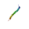
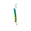
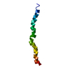
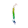
There are two copies of the biologically relevant assembly in the asymmetric unit, comprising chains B,C and D (copy 1) and E, G, and H (copy 2). The correct oligomerization size and orientation of chains was determined experimentally in solution. Chains A and F correspond to crystallographic packing artefacts.
Protocol: SINGLE WAVELENGTH / Monochromatic (M) / Laue (L): M / Scattering type: x-ray
Radiation wavelength
Wavelength: 0.9795 Å / Relative weight: 1
Reflection
Resolution: 1.8→32.1 Å / Num. obs: 31718 / % possible obs: 68.2 % / Redundancy: 2 % / Biso Wilson estimate: 32.35 Å2 / Rmerge(I) obs: 0.044 / Net I/σ(I): 11.3
Reflection shell
Resolution: 1.8→1.85 Å / Redundancy: 0.1 % / Rmerge(I) obs: 0.318 / Mean I/σ(I) obs: 3.9 / % possible all: 3.9
-
Processing
Software
Name
Version
Classification
BUSTER
2.10.2
refinement
XDS
datareduction
XSCALE
datascaling
PHENIX
phasing
Refinement
Method to determine structure: SAD / Resolution: 1.8→32.1 Å / Cor.coef. Fo:Fc: 0.9408 / Cor.coef. Fo:Fc free: 0.9244 / SU R Cruickshank DPI: 0.213 / Cross valid method: THROUGHOUT / σ(F): 0 / SU R Blow DPI: 0.227 / SU Rfree Blow DPI: 0.175 / SU Rfree Cruickshank DPI: 0.172
Rfactor
Num. reflection
% reflection
Selection details
Rfree
0.2392
1600
5.04 %
RANDOM
Rwork
0.2108
-
-
-
obs
0.2123
31718
67.84 %
-
Displacement parameters
Biso mean: 51.24 Å2
Baniso -1
Baniso -2
Baniso -3
1-
-6.2821 Å2
0 Å2
0 Å2
2-
-
1.2079 Å2
0 Å2
3-
-
-
5.0742 Å2
Refine analyze
Luzzati coordinate error obs: 0.3 Å
Refinement step
Cycle: LAST / Resolution: 1.8→32.1 Å
Protein
Nucleic acid
Ligand
Solvent
Total
Num. atoms
3647
0
25
249
3921
Refine LS restraints
Refine-ID
Type
Dev ideal
Number
Restraint function
Weight
X-RAY DIFFRACTION
t_bond_d
0.01
3708
HARMONIC
2
X-RAY DIFFRACTION
t_angle_deg
0.96
4984
HARMONIC
2
X-RAY DIFFRACTION
t_dihedral_angle_d
1455
SINUSOIDAL
2
X-RAY DIFFRACTION
t_incorr_chiral_ct
X-RAY DIFFRACTION
t_pseud_angle
X-RAY DIFFRACTION
t_trig_c_planes
170
HARMONIC
2
X-RAY DIFFRACTION
t_gen_planes
496
HARMONIC
5
X-RAY DIFFRACTION
t_it
3708
HARMONIC
20
X-RAY DIFFRACTION
t_nbd
X-RAY DIFFRACTION
t_omega_torsion
2.2
X-RAY DIFFRACTION
t_other_torsion
18.8
X-RAY DIFFRACTION
t_improper_torsion
X-RAY DIFFRACTION
t_chiral_improper_torsion
497
SEMIHARMONIC
5
X-RAY DIFFRACTION
t_sum_occupancies
X-RAY DIFFRACTION
t_utility_distance
X-RAY DIFFRACTION
t_utility_angle
X-RAY DIFFRACTION
t_utility_torsion
X-RAY DIFFRACTION
t_ideal_dist_contact
4521
SEMIHARMONIC
4
LS refinement shell
Resolution: 1.8→1.86 Å / Total num. of bins used: 16
Rfactor
Num. reflection
% reflection
Rfree
0.3087
12
6.49 %
Rwork
0.2103
173
-
all
0.2153
185
-
obs
-
-
4.35 %
Refinement TLS params.
Method: refined / Refine-ID: X-RAY DIFFRACTION
ID
L11 (°2)
L12 (°2)
L13 (°2)
L22 (°2)
L23 (°2)
L33 (°2)
S11 (Å °)
S12 (Å °)
S13 (Å °)
S21 (Å °)
S22 (Å °)
S23 (Å °)
S31 (Å °)
S32 (Å °)
S33 (Å °)
T11 (Å2)
T12 (Å2)
T13 (Å2)
T22 (Å2)
T23 (Å2)
T33 (Å2)
Origin x (Å)
Origin y (Å)
Origin z (Å)
1
3.1949
0.226
1.3692
2.0987
-1.8103
9.9416
-0.1545
0.3458
-0.3282
-0.021
0.0347
-0.1093
0.2702
0.1102
0.1197
-0.242
0.0497
0.0028
-0.1009
-0.0807
-0.0997
14.2946
21.281
43.9163
2
2.4401
0.7066
2.0089
2.4972
0.865
9.6432
-0.0183
-0.0074
-0.1079
0.2448
-0.0021
-0.1264
0.3561
0.1292
0.0204
-0.3511
0.0046
-0.0027
-0.2576
-0.0458
-0.2232
19.1268
26.841
57.8562
3
0.1287
-0.1509
-0.7322
0.7899
1.9626
11.969
-0.0239
-0.05
-0.0728
0.0275
0.054
0.0283
-0.0079
0.2437
-0.0301
-0.4014
0.0302
-0.0164
-0.148
-0.0637
-0.2069
29.1925
31.5397
59.434
4
1.0905
-0.0421
-0.3265
0.6864
1.4753
14.0407
0.0945
0.0285
0.0654
-0.0278
0.0862
-0.0274
-0.6281
-0.1857
-0.1807
-0.3592
0.0319
-0.0071
-0.2955
-0.0474
-0.2291
18.9409
35.1381
63.4761
5
2.4263
-0.1042
-2.1548
3.5569
0.3096
6.1472
-0.006
-0.1135
0.0701
-0.4031
0.1027
-0.1899
-0.6183
0.2015
-0.0967
-0.4281
-0.0402
0.0046
-0.1911
-0.0893
-0.2204
42.691
25.8986
30.2486
6
2.3804
-0.307
-0.3085
1.6323
-0.2824
9.352
-0.1514
-0.0776
0.3697
0.0162
-0.0554
-0.0665
-0.5922
0.6906
0.2068
-0.1335
-0.037
0.0163
-0.0472
-0.0082
-0.0836
37.6759
30.1012
45.4554
7
0.691
-0.2978
-0.8852
0.4164
0.2634
9.3625
0.0917
0.0083
-0.0178
-0.0323
0.1114
0.0384
0.2625
-0.0715
-0.2031
-0.2763
-0.0494
-0.0009
-0.2144
-0.0421
-0.1915
41.9516
18.5845
24.0675
8
0.1043
0.0262
2.0533
0.6463
2.1014
12.6869
-0.0264
-0.0899
0.0961
-0.0057
-0.0178
0.0383
0.1018
0.2253
0.0442
-0.315
-0.0406
0.0101
-0.0262
-0.0799
-0.1967
52.3323
21.0982
27.8978
Refinement TLS group
ID
Refine-ID
Refine TLS-ID
Selection details
1
X-RAY DIFFRACTION
1
{ A|129 - A|175 }
2
X-RAY DIFFRACTION
2
{ B|129 - B|179 }
3
X-RAY DIFFRACTION
3
{ C|123 - C|179 }
4
X-RAY DIFFRACTION
4
{ D|124 - D|179 }
5
X-RAY DIFFRACTION
5
{ E|127 - E|179 }
6
X-RAY DIFFRACTION
6
{ F|127 - F|176 }
7
X-RAY DIFFRACTION
7
{ G|124 - G|179 }
8
X-RAY DIFFRACTION
8
{ H|123 - H|179 }
+
About Yorodumi
-
News
-
Feb 9, 2022. New format data for meta-information of EMDB entries
New format data for meta-information of EMDB entries
Version 3 of the EMDB header file is now the official format.
The previous official version 1.9 will be removed from the archive.
In the structure databanks used in Yorodumi, some data are registered as the other names, "COVID-19 virus" and "2019-nCoV". Here are the details of the virus and the list of structure data.
Jan 31, 2019. EMDB accession codes are about to change! (news from PDBe EMDB page)
EMDB accession codes are about to change! (news from PDBe EMDB page)
The allocation of 4 digits for EMDB accession codes will soon come to an end. Whilst these codes will remain in use, new EMDB accession codes will include an additional digit and will expand incrementally as the available range of codes is exhausted. The current 4-digit format prefixed with “EMD-” (i.e. EMD-XXXX) will advance to a 5-digit format (i.e. EMD-XXXXX), and so on. It is currently estimated that the 4-digit codes will be depleted around Spring 2019, at which point the 5-digit format will come into force.
The EM Navigator/Yorodumi systems omit the EMD- prefix.
Related info.:Q: What is EMD? / ID/Accession-code notation in Yorodumi/EM Navigator
Yorodumi is a browser for structure data from EMDB, PDB, SASBDB, etc.
This page is also the successor to EM Navigator detail page, and also detail information page/front-end page for Omokage search.
The word "yorodu" (or yorozu) is an old Japanese word meaning "ten thousand". "mi" (miru) is to see.
Related info.:EMDB / PDB / SASBDB / Comparison of 3 databanks / Yorodumi Search / Aug 31, 2016. New EM Navigator & Yorodumi / Yorodumi Papers / Jmol/JSmol / Function and homology information / Changes in new EM Navigator and Yorodumi
 Movie
Movie Controller


 Open data
 Basic information
 Components
 Keywords
 Function and homology information
 X-RAY DIFFRACTION /
  Authors
 United Kingdom, 2items
  Citation
 Structure visualization
 Downloads & links
 Other downloads
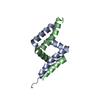

 PDBj
PDBj Assembly


 Mass: 94.971 Da / Num. of mol.: 2 / Source method: obtained synthetically / Formula: PO4
 Mass: 96.063 Da / Num. of mol.: 3 / Source method: obtained synthetically / Formula: SO4
 Mass: 18.015 Da / Num. of mol.: 249 / Source method: isolated from a natural source / Formula: H2O
 Sample preparation
 Processing