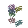
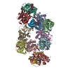
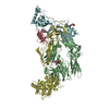
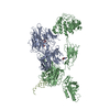
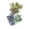
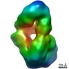
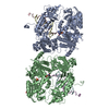
ジャーナル: Proc Natl Acad Sci U S A / 年: 2011 タイトル: Unique structure of iC3b resolved at a resolution of 24 Å by 3D-electron microscopy. 著者: Martin Alcorlo / Ruben Martínez-Barricarte / Francisco J Fernández / César Rodríguez-Gallego / Adam Round / M Cristina Vega / Claire L Harris / Santiago Rodríguez de Cordoba / Oscar Llorca / 要旨: Activation of C3, deposition of C3b on the target surface, and subsequent amplification by formation of a C3-cleaving enzyme (C3-convertase; C3bBb) triggers the effector functions of complement that ...Activation of C3, deposition of C3b on the target surface, and subsequent amplification by formation of a C3-cleaving enzyme (C3-convertase; C3bBb) triggers the effector functions of complement that result in inflammation and cell lysis. Concurrently, surface-bound C3b is proteolyzed to iC3b by factor I and appropriate cofactors. iC3b then interacts with the complement receptors (CR) of the Ig superfamily, CR2 (CD21), CR3 (CD11b/CD18), and CR4 (CD11c/CD18) on leukocytes, down-modulating inflammation, enhancing B cell-mediated immunity, and targeting pathogens for clearance by phagocytosis. Using EM and small-angle X-ray scattering, we now present a medium-resolution structure of iC3b (24 Å). iC3b displays a unique conformation with structural features distinct from any other C3 fragment. The macroglobulin ring in iC3b is similar to that in C3b, whereas the TED (thioester-containing domain) domain and the remnants of the CUB (complement protein subcomponents C1r/C1s, urchin embryonic growth factor and bone morphogenetic protein 1) domain have moved to locations more similar to where they were in native C3. A consequence of this large conformational change is the disruption of the factor B binding site, which renders iC3b unable to assemble a C3-convertase. This structural model also justifies the decreased interaction between iC3b and complement regulators and the recognition of iC3b by the CR of the Ig superfamily, CR2, CR3, and CR4. These data further illustrate the extraordinary conformational versatility of C3 to accommodate a great diversity of functional activities.
Legacy - 非点収差: Objective lens astigmatism was corrected at 80,000 times magnification
撮影
カテゴリ: FILM / フィルム・検出器のモデル: KODAK SO-163 FILM / デジタル化 - スキャナー: OTHER / デジタル化 - サンプリング間隔: 10.5 µm / 実像数: 112 / 平均電子線量: 10 e/Å2 詳細: Images scanned with Minolta Dimage Scan Multi Pro scanner at 2400 dpi and averaged to a final 4.2 Angstroms pixel at the specimen ビット/ピクセル: 16
 ムービー
ムービー コントローラー


 データを開く
 基本情報
基本情報 マップデータ
 試料
試料 キーワード
キーワード Homo sapiens (ヒト)
 データ登録者
 引用
引用
 構造の表示
構造の表示 ムービービューア
 UCSF Chimera
 ダウンロードとリンク
 http://ftp.pdbj.org/pub/emdb/structures/EMD-1908
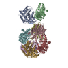
 Z (Sec.)
 Y (Row.)
 X (Col.)


 試料の構成要素
 解析
解析 電子顕微鏡法
