 Movie
Movie Controller

[English] 日本語
 Yorodumi
- PDB-3l8b: Crystal structure of a replicative DNA polymerase bound to the ox... -
+ Open data

- Basic information
| Entry | Database: PDB / ID: 3l8b | ||||||
|---|---|---|---|---|---|---|---|
| Title | Crystal structure of a replicative DNA polymerase bound to the oxidized guanine lesion guanidinohydantoin | ||||||
 Components Components |
| ||||||
 Keywords Keywords | transferase/DNA / DNA polymerase RB69 gp43 / protein-DNA complex / oxidative DNA lesion / guanidinohydantoin / DNA replication / DNA-binding / DNA-directed DNA polymerase / Exonuclease / Hydrolase / Nuclease / Nucleotidyltransferase / Transferase / transferase-DNA complex | ||||||
| Function / homology |  Function and homology information Function and homology informationbidirectional double-stranded viral DNA replication / Hydrolases; Acting on ester bonds; Exodeoxyribonucleases producing 5'-phosphomonoesters / 3'-5' exonuclease activity / DNA-templated DNA replication / DNA-directed DNA polymerase / DNA-directed DNA polymerase activity / nucleotide binding / DNA binding / metal ion binding Similarity search - Function | ||||||
| Biological species |  Enterobacteria phage RB69 (virus) Enterobacteria phage RB69 (virus) | ||||||
| Method |  X-RAY DIFFRACTION / SYNCHROTRON / MOLECULAR REPLACEMENT / Resolution: 2.15 Å X-RAY DIFFRACTION / SYNCHROTRON / MOLECULAR REPLACEMENT / Resolution: 2.15 Å | ||||||
 Authors Authors | Aller, P. / Ye, Y. / Wallace, S.S. / Burrows, C.J. / Doublie, S. | ||||||
 Citation Citation | Journal: Biochemistry / Year: 2010 Title: Crystal structure of a replicative DNA polymerase bound to the oxidized guanine lesion guanidinohydantoin. Authors: Aller, P. / Ye, Y. / Wallace, S.S. / Burrows, C.J. / Doublie, S. | ||||||
| History |
|
- Structure visualization
| Structure viewer | Molecule: MolmilJmol/JSmol |
|---|
- Downloads & links
-Download
| PDBx/mmCIF format | 3l8b.cif.gz | 423.1 KB | Display | PDBx/mmCIF format |
|---|---|---|---|---|
| PDB format | pdb3l8b.ent.gz | 334.9 KB | Display | PDB format |
| PDBx/mmJSON format | 3l8b.json.gz | Tree view | PDBx/mmJSON format | |
| Others |  Other downloads Other downloads |
-Validation report
| Arichive directory | https://data.pdbj.org/pub/pdb/validation_reports/l8/3l8bftp://data.pdbj.org/pub/pdb/validation_reports/l8/3l8b | HTTPS FTP |
|---|
-Related structure data
| Related structure data | 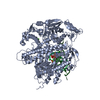 1ig9S S: Starting model for refinement |
|---|---|
| Similar structure data |


-Links
 PDBj
PDBj


- Assembly
| Deposited unit | 
| ||||||||
|---|---|---|---|---|---|---|---|---|---|
| 1 | 
| ||||||||
| 2 | 
| ||||||||
| Unit cell |
|
-Components
| #1: Protein | Mass: 105069.586 Da / Num. of mol.: 2 / Mutation: D222A, D327A Source method: isolated from a genetically manipulated source Source: (gene. exp.) Enterobacteria phage RB69 (virus) / Gene: 43, gp43 / Production host:  #2: DNA chain | Mass: 5522.583 Da / Num. of mol.: 2 / Source method: obtained synthetically / Details: DNA template #3: DNA chain | Mass: 3991.610 Da / Num. of mol.: 2 / Source method: obtained synthetically / Details: DNA primer #4: Chemical |   Mass: 96.063 Da / Num. of mol.: 2 / Source method: obtained synthetically / Formula: SO4 Mass: 96.063 Da / Num. of mol.: 2 / Source method: obtained synthetically / Formula: SO4#5: Water | ChemComp-HOH / |  Mass: 18.015 Da / Num. of mol.: 998 / Source method: isolated from a natural source / Formula: H2O Mass: 18.015 Da / Num. of mol.: 998 / Source method: isolated from a natural source / Formula: H2O |
|---|
-Experimental details
-Experiment
| Experiment | Method: X-RAY DIFFRACTION / Number of used crystals: 1 |
|---|
- Sample preparation
| Crystal | Density Matthews: 2.81 Å3/Da / Density % sol: 56.19 % Description: The Structure Factor file contains Friedel pairs |
|---|---|
| Crystal grow | Temperature: 297 K / Method: vapor diffusion, hanging drop / pH: 7 Details: reservoir: 12% PEG2000MME, 100mM Sodium Acetate, 150mM Magnesium Sulfate, 100mM Hepes pH 7.0, 6% Glycerol and 2mM Beta-mercaptoethanol, VAPOR DIFFUSION, HANGING DROP, temperature 297K |
-Data collection
| Diffraction | Mean temperature: 100 K |
|---|---|
| Diffraction source | Source: SYNCHROTRON / Site: APS  / Beamline: 23-ID-B / Wavelength: 1.0332 Å / Beamline: 23-ID-B / Wavelength: 1.0332 Å |
| Detector | Type: MARMOSAIC 300 mm CCD / Detector: CCD / Date: Oct 23, 2008 Details: Si(111) Double Crystal Monochrometer. Adjustable focusing mirrors in K-B geometry |
| Radiation | Monochromator: Double crystal cryo-cooled Si(111) / Protocol: SINGLE WAVELENGTH / Monochromatic (M) / Laue (L): M / Scattering type: x-ray |
| Radiation wavelength | Wavelength: 1.0332 Å / Relative weight: 1 |
| Reflection | Resolution: 2.15→48.81 Å / Num. all: 270208 / Num. obs: 239932 / % possible obs: 88.8 % / Observed criterion σ(I): 0 / Redundancy: 4.3 % / Biso Wilson estimate: 30.1 Å2 / Rmerge(I) obs: 0.126 / Net I/σ(I): 9.9 |
| Reflection shell | Resolution: 2.15→2.23 Å / Redundancy: 4 % / Rmerge(I) obs: 0.707 / Mean I/σ(I) obs: 2 / % possible all: 99.5 |
- Processing
| Software |
| |||||||||||||||||||||||||
|---|---|---|---|---|---|---|---|---|---|---|---|---|---|---|---|---|---|---|---|---|---|---|---|---|---|---|
| Refinement | Method to determine structure: MOLECULAR REPLACEMENT Starting model: PDB entry 1IG9 Resolution: 2.15→48.81 Å / Isotropic thermal model: anisotropic / σ(F): 0 / Stereochemistry target values: Engh & Huber
| |||||||||||||||||||||||||
| Displacement parameters |
| |||||||||||||||||||||||||
| Refine analyze | Luzzati coordinate error obs: 0.28 Å / Luzzati d res low obs: 5 Å / Luzzati sigma a obs: 0.33 Å | |||||||||||||||||||||||||
| Refinement step | Cycle: LAST / Resolution: 2.15→48.81 Å
| |||||||||||||||||||||||||
| Refine LS restraints |
|
