- PDB-2p5o: Crystal structure of RB69 GP43 in complex with DNA containing an ... -
+
Open data
ID or keywords:
Loading...
-
Basic information
Entry
Database: PDB / ID: 2p5o
Title
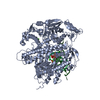
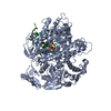
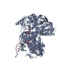
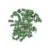
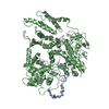
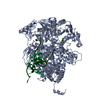
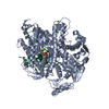
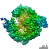
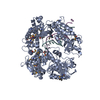
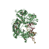
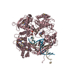
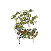
Crystal structure of RB69 GP43 in complex with DNA containing an abasic site analog
Components
DNA polymerase
Primer DNA
Template DNA
Keywords
transferase/DNA / DNA POLYMERASE / ABASIC SITE / DNA LESION / EXONUCLEASE SWITCH / transferase-DNA COMPLEX
Function / homology
Function and homology information
bidirectional double-stranded viral DNA replication / Hydrolases; Acting on ester bonds; Exodeoxyribonucleases producing 5'-phosphomonoesters / 3'-5' exonuclease activity / DNA-templated DNA replication / DNA-directed DNA polymerase / DNA-directed DNA polymerase activity / nucleotide binding / DNA binding / metal ion binding Similarity search - Function
DnaQ-like 3'-5' exonuclease / Monooxygenase - #300 / Ribonuclease H-like motif / DNA-directed DNA polymerase T4 type / DNA Polymerase; Chain A, domain 1 / DNA Polymerase, chain B, domain 1 / B family DNA polymerase, finger domain / Palm domain of DNA polymerase / B family DNA polymerase, palm domain / Monooxygenase ...DnaQ-like 3'-5' exonuclease / Monooxygenase - #300 / Ribonuclease H-like motif / DNA-directed DNA polymerase T4 type / DNA Polymerase; Chain A, domain 1 / DNA Polymerase, chain B, domain 1 / B family DNA polymerase, finger domain / Palm domain of DNA polymerase / B family DNA polymerase, palm domain / Monooxygenase / : / DNA-directed DNA polymerase, family B, multifunctional domain / DNA-directed DNA polymerase, family B, conserved site / DNA polymerase family B signature. / DNA polymerase family B / DNA polymerase family B, exonuclease domain / DNA-directed DNA polymerase, family B, exonuclease domain / DNA polymerase, palm domain superfamily / DNA polymerase type-B family / DNA-directed DNA polymerase, family B / Ribonuclease H-like superfamily/Ribonuclease H / Nucleotidyltransferase; domain 5 / Helix Hairpins / Ribonuclease H superfamily / Ribonuclease H-like superfamily / DNA/RNA polymerase superfamily / Alpha-Beta Complex / Up-down Bundle / 2-Layer Sandwich / Orthogonal Bundle / 3-Layer(aba) Sandwich / Mainly Alpha / Alpha Beta Similarity search - Domain/homology
E: Template DNA F: Primer DNA G: Template DNA H: Primer DNA I: Template DNA J: Primer DNA K: Template DNA L: Primer DNA A: DNA polymerase B: DNA polymerase C: DNA polymerase D: DNA polymerase
In the structure databanks used in Yorodumi, some data are registered as the other names, "COVID-19 virus" and "2019-nCoV". Here are the details of the virus and the list of structure data.
Jan 31, 2019. EMDB accession codes are about to change! (news from PDBe EMDB page)
EMDB accession codes are about to change! (news from PDBe EMDB page)
The allocation of 4 digits for EMDB accession codes will soon come to an end. Whilst these codes will remain in use, new EMDB accession codes will include an additional digit and will expand incrementally as the available range of codes is exhausted. The current 4-digit format prefixed with “EMD-” (i.e. EMD-XXXX) will advance to a 5-digit format (i.e. EMD-XXXXX), and so on. It is currently estimated that the 4-digit codes will be depleted around Spring 2019, at which point the 5-digit format will come into force.
The EM Navigator/Yorodumi systems omit the EMD- prefix.
Related info.:Q: What is EMD? / ID/Accession-code notation in Yorodumi/EM Navigator
Yorodumi is a browser for structure data from EMDB, PDB, SASBDB, etc.
This page is also the successor to EM Navigator detail page, and also detail information page/front-end page for Omokage search.
The word "yorodu" (or yorozu) is an old Japanese word meaning "ten thousand". "mi" (miru) is to see.
Related info.:EMDB / PDB / SASBDB / Comparison of 3 databanks / Yorodumi Search / Aug 31, 2016. New EM Navigator & Yorodumi / Yorodumi Papers / Jmol/JSmol / Function and homology information / Changes in new EM Navigator and Yorodumi
 Movie
Movie Controller

 Yorodumi
 Open data
 Basic information
 Components
 Keywords
 Function and homology information
 Enterobacteria phage RB69 (virus)
 X-RAY DIFFRACTION /
  Authors
 Citation
 Structure visualization
 Downloads & links
 Other downloads
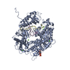


 PDBj
PDBj


 Assembly
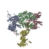

 Mass: 18.015 Da / Num. of mol.: 495 / Source method: isolated from a natural source / Formula: H2O
 Sample preparation
 / Beamline: X12C / Wavelength: 0.9500, 0.9788, 0.9790
 Processing