 Movie
Movie Controller

[English] 日本語
 Yorodumi
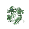
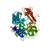
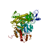
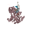
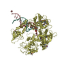
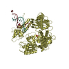
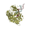
- PDB-1clq: CRYSTAL STRUCTURE OF A REPLICATION FORK DNA POLYMERASE EDITING CO... -
+ Open data

- Basic information
| Entry | Database: PDB / ID: 1clq | ||||||
|---|---|---|---|---|---|---|---|
| Title | CRYSTAL STRUCTURE OF A REPLICATION FORK DNA POLYMERASE EDITING COMPLEX AT 2.7 A RESOLUTION | ||||||
 Components Components |
| ||||||
 Keywords Keywords | TRANSFERASE/DNA / DNA POLYMERASE / GP43 / PROOFREADING / EDITING / REPLICATION / TRANSFERASE-DNA COMPLEX | ||||||
| Function / homology |  Function and homology information Function and homology informationbidirectional double-stranded viral DNA replication / Hydrolases; Acting on ester bonds; Exodeoxyribonucleases producing 5'-phosphomonoesters / 3'-5' exonuclease activity / DNA-templated DNA replication / DNA-directed DNA polymerase / DNA-directed DNA polymerase activity / nucleotide binding / DNA binding / metal ion binding Similarity search - Function | ||||||
| Biological species |  Enterobacteria phage RB69 (virus) Enterobacteria phage RB69 (virus) | ||||||
| Method |  X-RAY DIFFRACTION / SYNCHROTRON / MOLECULAR REPLACEMENT / Resolution: 2.7 Å X-RAY DIFFRACTION / SYNCHROTRON / MOLECULAR REPLACEMENT / Resolution: 2.7 Å | ||||||
 Authors Authors | Shamoo, Y. / Steitz, T.A. | ||||||
 Citation Citation | Journal: Cell(Cambridge,Mass.) / Year: 1999 Title: Building a replisome from interacting pieces: sliding clamp complexed to a peptide from DNA polymerase and a polymerase editing complex. Authors: Shamoo, Y. / Steitz, T.A. | ||||||
| History |
|
- Structure visualization
| Structure viewer | Molecule: MolmilJmol/JSmol |
|---|
- Downloads & links
-Download
| PDBx/mmCIF format | 1clq.cif.gz | 211.2 KB | Display | PDBx/mmCIF format |
|---|---|---|---|---|
| PDB format | pdb1clq.ent.gz | 169.5 KB | Display | PDB format |
| PDBx/mmJSON format | 1clq.json.gz | Tree view | PDBx/mmJSON format | |
| Others |  Other downloads Other downloads |
-Validation report
| Arichive directory | https://data.pdbj.org/pub/pdb/validation_reports/cl/1clqftp://data.pdbj.org/pub/pdb/validation_reports/cl/1clq | HTTPS FTP |
|---|
-Related structure data
| Related structure data |  1b77C  1b8hC  1wajS S: Starting model for refinement C: citing same article ( |
|---|---|
| Similar structure data |


-Links
 PDBj
PDBj


- Assembly
| Deposited unit | 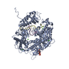
| ||||||||||
|---|---|---|---|---|---|---|---|---|---|---|---|
| 1 |
| ||||||||||
| Unit cell |
|
-Components
-DNA chain , 2 types, 2 molecules ED
| #1: DNA chain | Mass: 3358.211 Da / Num. of mol.: 1 / Source method: obtained synthetically / Details: PRIMER-TEMPLATE DNA |
|---|---|
| #2: DNA chain | Mass: 3678.403 Da / Num. of mol.: 1 / Source method: obtained synthetically |
-Protein , 1 types, 1 molecules A
| #3: Protein | Mass: 104743.156 Da / Num. of mol.: 1 / Fragment: RESIDUES 1-903 Source method: isolated from a genetically manipulated source Source: (gene. exp.) Enterobacteria phage RB69 (virus) / Genus: T4-like viruses / Gene: GENE 43 / Cell line (production host): BL21(DE3) / Production host:  |
|---|
-Non-polymers , 3 types, 231 molecules 


| #4: Chemical | ChemComp-CA /  Mass: 40.078 Da / Num. of mol.: 9 / Source method: obtained synthetically / Formula: Ca Mass: 40.078 Da / Num. of mol.: 9 / Source method: obtained synthetically / Formula: Ca#5: Chemical | ChemComp-GDP / |  Type: RNA linking / Mass: 443.201 Da / Num. of mol.: 1 / Source method: obtained synthetically / Formula: C10H15N5O11P2 / Comment: GDP, energy-carrying molecule*YM Type: RNA linking / Mass: 443.201 Da / Num. of mol.: 1 / Source method: obtained synthetically / Formula: C10H15N5O11P2 / Comment: GDP, energy-carrying molecule*YM#6: Water | ChemComp-HOH / | Mass: 18.015 Da / Num. of mol.: 221 / Source method: isolated from a natural source / Formula: H2O |
|---|
-Experimental details
-Experiment
| Experiment | Method: X-RAY DIFFRACTION / Number of used crystals: 1 |
|---|
- Sample preparation
| Crystal | Density Matthews: 4.7 Å3/Da / Density % sol: 70.4 % | |||||||||||||||||||||||||
|---|---|---|---|---|---|---|---|---|---|---|---|---|---|---|---|---|---|---|---|---|---|---|---|---|---|---|
| Crystal grow | Temperature: 285 K / Method: vapor diffusion, hanging drop / pH: 6.5 Details: 12% (W/V) PEG 350MME, 150 MM CACL2, 100 MM SODIUM CACODYLATE, PH 6.5, 12 DEGREES C, VAPOR DIFFUSION, HANGING DROP, temperature 285K | |||||||||||||||||||||||||
| Components of the solutions |
| |||||||||||||||||||||||||
| Crystal grow | *PLUS Temperature: 12 ℃ | |||||||||||||||||||||||||
| Components of the solutions | *PLUS
|
-Data collection
| Diffraction | Mean temperature: 100 K |
|---|---|
| Diffraction source | Source: SYNCHROTRON / Site: CHESS  / Beamline: A1 / Wavelength: 0.9188 / Beamline: A1 / Wavelength: 0.9188 |
| Detector | Type: ADSC / Detector: CCD / Date: Oct 15, 1998 / Details: SILICON 111 BENDING MIRROR |
| Radiation | Protocol: SINGLE WAVELENGTH / Monochromatic (M) / Laue (L): M / Scattering type: x-ray |
| Radiation wavelength | Wavelength: 0.9188 Å / Relative weight: 1 |
| Reflection | Resolution: 2.7→40 Å / Num. obs: 51455 / % possible obs: 97.2 % / Observed criterion σ(I): 2 / Redundancy: 2.2 % / Biso Wilson estimate: 50 Å2 / Rsym value: 0.08 / Net I/σ(I): 18.1 |
| Reflection | *PLUS Lowest resolution: 40 Å / Num. obs: 23329 / Num. measured all: 51455 / Rmerge(I) obs: 0.08 |
- Processing
| Software |
| ||||||||||||||||||||||||||||||||||||||||||||||||||||||||||||||||||||||||||||||||
|---|---|---|---|---|---|---|---|---|---|---|---|---|---|---|---|---|---|---|---|---|---|---|---|---|---|---|---|---|---|---|---|---|---|---|---|---|---|---|---|---|---|---|---|---|---|---|---|---|---|---|---|---|---|---|---|---|---|---|---|---|---|---|---|---|---|---|---|---|---|---|---|---|---|---|---|---|---|---|---|---|---|
| Refinement | Method to determine structure: MOLECULAR REPLACEMENT Starting model: 1WAJ Resolution: 2.7→40 Å / Rfactor Rfree error: 0.004 / Data cutoff high rms absF: 505175.56 / Isotropic thermal model: RESTRAINED / Cross valid method: THROUGHOUT / σ(F): 0
| ||||||||||||||||||||||||||||||||||||||||||||||||||||||||||||||||||||||||||||||||
| Solvent computation | Solvent model: FLAT MODEL / Bsol: 46.43 Å2 / ksol: 0.31 e/Å3 | ||||||||||||||||||||||||||||||||||||||||||||||||||||||||||||||||||||||||||||||||
| Displacement parameters | Biso mean: 58.4 Å2
| ||||||||||||||||||||||||||||||||||||||||||||||||||||||||||||||||||||||||||||||||
| Refine analyze |
| ||||||||||||||||||||||||||||||||||||||||||||||||||||||||||||||||||||||||||||||||
| Refinement step | Cycle: LAST / Resolution: 2.7→40 Å
| ||||||||||||||||||||||||||||||||||||||||||||||||||||||||||||||||||||||||||||||||
| Refine LS restraints |
| ||||||||||||||||||||||||||||||||||||||||||||||||||||||||||||||||||||||||||||||||
| LS refinement shell | Resolution: 2.7→2.87 Å / Rfactor Rfree error: 0.014 / Total num. of bins used: 6
| ||||||||||||||||||||||||||||||||||||||||||||||||||||||||||||||||||||||||||||||||
| Xplor file |
| ||||||||||||||||||||||||||||||||||||||||||||||||||||||||||||||||||||||||||||||||
| Software | *PLUS Name: CNS / Version: 0.4 / Classification: refinement | ||||||||||||||||||||||||||||||||||||||||||||||||||||||||||||||||||||||||||||||||
| Refinement | *PLUS Highest resolution: 2.7 Å / σ(F): 0 / % reflection Rfree: 9.7 % / Rfactor obs: 0.23 / Rfactor Rwork: 0.23 | ||||||||||||||||||||||||||||||||||||||||||||||||||||||||||||||||||||||||||||||||
| Solvent computation | *PLUS | ||||||||||||||||||||||||||||||||||||||||||||||||||||||||||||||||||||||||||||||||
| Displacement parameters | *PLUS Biso mean: 58.4 Å2 | ||||||||||||||||||||||||||||||||||||||||||||||||||||||||||||||||||||||||||||||||
| Refine LS restraints | *PLUS
| ||||||||||||||||||||||||||||||||||||||||||||||||||||||||||||||||||||||||||||||||
| LS refinement shell | *PLUS Rfactor Rfree: 0.399 / % reflection Rfree: 10.8 % / Rfactor Rwork: 0.347 |
