 Movie
Movie Controller

[English] 日本語
 Yorodumi
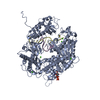
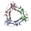
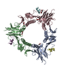
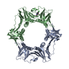
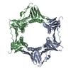
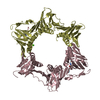
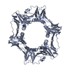
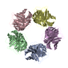
- PDB-1b77: BUILDING A REPLISOME STRUCTURE FROM INTERACTING PIECES: A SLIDING... -
+ Open data

- Basic information
| Entry | Database: PDB / ID: 1b77 | ||||||
|---|---|---|---|---|---|---|---|
| Title | BUILDING A REPLISOME STRUCTURE FROM INTERACTING PIECES: A SLIDING CLAMP COMPLEXED WITH AN INTERACTION PEPTIDE FROM DNA POLYMERASE | ||||||
 Components Components | PROTEIN (SLIDING CLAMP) | ||||||
 Keywords Keywords | SLIDING CLAMP / GP45 / REPLISOME | ||||||
| Function / homology |  Function and homology information Function and homology informationviral DNA genome replication / DNA polymerase processivity factor activity / viral transcription / DNA replication Similarity search - Function | ||||||
| Biological species |  Enterobacteria phage RB69 (virus) Enterobacteria phage RB69 (virus) | ||||||
| Method |  X-RAY DIFFRACTION / SYNCHROTRON / MOLECULAR REPLACEMENT / Resolution: 2.1 Å X-RAY DIFFRACTION / SYNCHROTRON / MOLECULAR REPLACEMENT / Resolution: 2.1 Å | ||||||
 Authors Authors | Shamoo, Y. / Steitz, T.A. | ||||||
 Citation Citation | Journal: Cell(Cambridge,Mass.) / Year: 1999 Title: Building a replisome from interacting pieces: sliding clamp complexed to a peptide from DNA polymerase and a polymerase editing complex. Authors: Shamoo, Y. / Steitz, T.A. | ||||||
| History |
|
- Structure visualization
| Structure viewer | Molecule: MolmilJmol/JSmol |
|---|
- Downloads & links
-Download
| PDBx/mmCIF format | 1b77.cif.gz | 153.2 KB | Display | PDBx/mmCIF format |
|---|---|---|---|---|
| PDB format | pdb1b77.ent.gz | 122.6 KB | Display | PDB format |
| PDBx/mmJSON format | 1b77.json.gz | Tree view | PDBx/mmJSON format | |
| Others |  Other downloads Other downloads |
-Validation report
| Arichive directory | https://data.pdbj.org/pub/pdb/validation_reports/b7/1b77ftp://data.pdbj.org/pub/pdb/validation_reports/b7/1b77 | HTTPS FTP |
|---|
-Related structure data


-Links
 PDBj
PDBj
- Assembly
| Deposited unit | 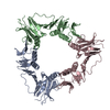
| ||||||||||||
|---|---|---|---|---|---|---|---|---|---|---|---|---|---|
| 1 |
| ||||||||||||
| Unit cell |
| ||||||||||||
| Noncrystallographic symmetry (NCS) | NCS oper:
|
-Components
| #1: Protein | Mass: 25133.475 Da / Num. of mol.: 3 Source method: isolated from a genetically manipulated source Source: (gene. exp.) Enterobacteria phage RB69 (virus) / Genus: T4-like viruses / Gene: GENE 45 / Cell line (production host): BL21(DE3) / Production host:  #2: Water | ChemComp-HOH / |  Mass: 18.015 Da / Num. of mol.: 713 / Source method: isolated from a natural source / Formula: H2O Mass: 18.015 Da / Num. of mol.: 713 / Source method: isolated from a natural source / Formula: H2O |
|---|
-Experimental details
-Experiment
| Experiment | Method: X-RAY DIFFRACTION / Number of used crystals: 1 |
|---|
- Sample preparation
| Crystal | Density Matthews: 2.8 Å3/Da / Density % sol: 47 % | ||||||||||||||||||||||||||||||||||||||||
|---|---|---|---|---|---|---|---|---|---|---|---|---|---|---|---|---|---|---|---|---|---|---|---|---|---|---|---|---|---|---|---|---|---|---|---|---|---|---|---|---|---|
| Crystal grow | pH: 7.5 / Details: 16-18% PEG 8000, 0.2 M CA ACETATE PH7.5 | ||||||||||||||||||||||||||||||||||||||||
| Crystal grow | *PLUS Temperature: 20 ℃ / Method: vapor diffusion | ||||||||||||||||||||||||||||||||||||||||
| Components of the solutions | *PLUS
|
-Data collection
| Diffraction | Mean temperature: 100 K |
|---|---|
| Diffraction source | Source: SYNCHROTRON / Site: CHESS  / Beamline: A1 / Wavelength: 0.9188 / Beamline: A1 / Wavelength: 0.9188 |
| Detector | Type: ADSC / Detector: CCD / Date: Nov 1, 1997 / Details: SILICON 111 BENDING MIRROR |
| Radiation | Protocol: SINGLE WAVELENGTH / Monochromatic (M) / Laue (L): M / Scattering type: x-ray |
| Radiation wavelength | Wavelength: 0.9188 Å / Relative weight: 1 |
| Reflection | Resolution: 2.1→20 Å / Num. obs: 47361 / % possible obs: 94.9 % / Observed criterion σ(I): 2 / Redundancy: 3.2 % / Biso Wilson estimate: 15 Å2 / Rsym value: 0.07 / Net I/σ(I): 6.6 |
| Reflection | *PLUS Num. measured all: 151555 / Rmerge(I) obs: 0.07 |
- Processing
| Software |
| ||||||||||||||||||||||||||||||||||||||||||||||||||||||||||||||||||||||||||||||||
|---|---|---|---|---|---|---|---|---|---|---|---|---|---|---|---|---|---|---|---|---|---|---|---|---|---|---|---|---|---|---|---|---|---|---|---|---|---|---|---|---|---|---|---|---|---|---|---|---|---|---|---|---|---|---|---|---|---|---|---|---|---|---|---|---|---|---|---|---|---|---|---|---|---|---|---|---|---|---|---|---|---|
| Refinement | Method to determine structure: MOLECULAR REPLACEMENT Starting model: PRELIMINARY COORDINATE OF T4 GP45 FROM J. KURIYAN Resolution: 2.1→20 Å / Rfactor Rfree error: 0.004 / Data cutoff high rms absF: 347237.93 / Isotropic thermal model: RESTRAINED / Cross valid method: THROUGHOUT / σ(F): 0
| ||||||||||||||||||||||||||||||||||||||||||||||||||||||||||||||||||||||||||||||||
| Solvent computation | Solvent model: FLAT MODEL / Bsol: 45.02 Å2 / ksol: 0.316 e/Å3 | ||||||||||||||||||||||||||||||||||||||||||||||||||||||||||||||||||||||||||||||||
| Displacement parameters | Biso mean: 32.4 Å2
| ||||||||||||||||||||||||||||||||||||||||||||||||||||||||||||||||||||||||||||||||
| Refine analyze |
| ||||||||||||||||||||||||||||||||||||||||||||||||||||||||||||||||||||||||||||||||
| Refinement step | Cycle: LAST / Resolution: 2.1→20 Å
| ||||||||||||||||||||||||||||||||||||||||||||||||||||||||||||||||||||||||||||||||
| Refine LS restraints |
| ||||||||||||||||||||||||||||||||||||||||||||||||||||||||||||||||||||||||||||||||
| LS refinement shell | Resolution: 2.1→2.23 Å / Rfactor Rfree error: 0.014 / Total num. of bins used: 6
| ||||||||||||||||||||||||||||||||||||||||||||||||||||||||||||||||||||||||||||||||
| Xplor file |
| ||||||||||||||||||||||||||||||||||||||||||||||||||||||||||||||||||||||||||||||||
| Software | *PLUS Name: CNS / Version: 0.4 / Classification: refinement | ||||||||||||||||||||||||||||||||||||||||||||||||||||||||||||||||||||||||||||||||
| Refine LS restraints | *PLUS
|
