 Movie
Movie Controller

[English] 日本語
 Yorodumi
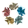
- PDB-1lj7: Crystal structure of calcium-depleted human C-reactive protein fr... -
+ Open data

- Basic information
| Entry | Database: PDB / ID: 1lj7 | ||||||
|---|---|---|---|---|---|---|---|
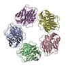
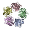
| Title | Crystal structure of calcium-depleted human C-reactive protein from perfectly twinned data | ||||||
 Components Components | C-reactive protein | ||||||
 Keywords Keywords | UNKNOWN FUNCTION / pentraxin fold / pentamer / decamer / twinned | ||||||
| Function / homology |  Function and homology information Function and homology informationregulation of interleukin-8 production / complement component C1q complex binding / opsonization / low-density lipoprotein particle binding / vasoconstriction / choline binding / negative regulation of mononuclear cell proliferation / Classical antibody-mediated complement activation / low-density lipoprotein particle receptor binding / negative regulation of macrophage derived foam cell differentiation ...regulation of interleukin-8 production / complement component C1q complex binding / opsonization / low-density lipoprotein particle binding / vasoconstriction / choline binding / negative regulation of mononuclear cell proliferation / Classical antibody-mediated complement activation / low-density lipoprotein particle receptor binding / negative regulation of macrophage derived foam cell differentiation / negative regulation of lipid storage / positive regulation of superoxide anion generation / acute-phase response / defense response to Gram-positive bacterium / inflammatory response / innate immune response / calcium ion binding / positive regulation of gene expression / : / extracellular region / identical protein binding Similarity search - Function | ||||||
| Biological species |  Homo sapiens (human) Homo sapiens (human) | ||||||
| Method |  X-RAY DIFFRACTION / SYNCHROTRON / MOLECULAR REPLACEMENT / Resolution: 3.15 Å X-RAY DIFFRACTION / SYNCHROTRON / MOLECULAR REPLACEMENT / Resolution: 3.15 Å | ||||||
 Authors Authors | Ramadan, M.A. / Shrive, A.K. / Holden, D. / Myles, D.A. / Volanakis, J.E. / DeLucas, L.J. / Greenhough, T.J. | ||||||
 Citation Citation | Journal: Acta Crystallogr.,Sect.D / Year: 2002 Title: The three-dimensional structure of calcium-depleted human C-reactive protein from perfectly twinned crystals. Authors: Ramadan, M.A. / Shrive, A.K. / Holden, D. / Myles, D.A. / Volanakis, J.E. / DeLucas, L.J. / Greenhough, T.J. | ||||||
| History |
| ||||||
| Remark 11 | A few small regions of the structure are not well defined due to the twinning and asociated ... A few small regions of the structure are not well defined due to the twinning and asociated disorder. The twinning 2-fold is parallel to the 2-fold ncs operator relating the two pentamers in the assymetric unit. Removal of calcium results in residues in the calcium- binding loop 140-150 becoming disordered and mobile. The loops and parts of loops that are visible are held in place by crystal contacts | ||||||
| Remark 300 | Biomolecule: 1,2 This entry contains the crystallographic assymetric unit which consists of 10 ... Biomolecule: 1,2 This entry contains the crystallographic assymetric unit which consists of 10 chain(s). See remark 350 for information on generating the biological molecule(s). It is, however, not clear whether the biomolecule consists of one or two pentameters |
- Structure visualization
| Structure viewer | Molecule: MolmilJmol/JSmol |
|---|
- Downloads & links
-Download
| PDBx/mmCIF format | 1lj7.cif.gz | 386.3 KB | Display | PDBx/mmCIF format |
|---|---|---|---|---|
| PDB format | pdb1lj7.ent.gz | 322.1 KB | Display | PDB format |
| PDBx/mmJSON format | 1lj7.json.gz | Tree view | PDBx/mmJSON format | |
| Others |  Other downloads Other downloads |
-Validation report
| Arichive directory | https://data.pdbj.org/pub/pdb/validation_reports/lj/1lj7ftp://data.pdbj.org/pub/pdb/validation_reports/lj/1lj7 | HTTPS FTP |
|---|
-Related structure data
| Related structure data | 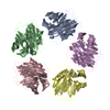 1gnhS S: Starting model for refinement |
|---|---|
| Similar structure data |

-Links
 PDBj
PDBj

- Assembly
| Deposited unit | 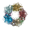
| ||||||||
|---|---|---|---|---|---|---|---|---|---|
| 1 | 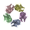
| ||||||||
| 2 | 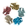
| ||||||||
| Unit cell |
|
-Components
| #1: Protein | Mass: 23068.039 Da / Num. of mol.: 10 / Source method: isolated from a natural source / Details: purified from serum / Source: (natural) Homo sapiens (human) / References: UniProt: P02741Has protein modification | Y | |
|---|
-Experimental details
-Experiment
| Experiment | Method: X-RAY DIFFRACTION / Number of used crystals: 17 |
|---|
- Sample preparation
| Crystal | Density Matthews: 3.6 Å3/Da / Density % sol: 66 % Description: Data in this section (200) refers to the twinned space group P4(1)22 |
|---|---|
| Crystal grow | Method: vapor diffusion, hanging drop / Details: VAPOR DIFFUSION, HANGING DROP |
| Crystal grow | *PLUS Details: DeLucas, L.J., (1987) J. Mol. Biol., 196, 741. |
-Data collection
| Diffraction | Mean temperature: 298 K |
|---|---|
| Diffraction source | Source: SYNCHROTRON / Site: SRS  / Beamline: PX7.2 / Wavelength: 1.488 Å / Beamline: PX7.2 / Wavelength: 1.488 Å |
| Detector | Type: CEA / Detector: FILM / Date: Jan 1, 1989 |
| Radiation | Monochromator: Germanium / Protocol: SINGLE WAVELENGTH / Monochromatic (M) / Laue (L): M / Scattering type: x-ray |
| Radiation wavelength | Wavelength: 1.488 Å / Relative weight: 1 |
| Reflection | Resolution: 3.15→72.6 Å / Num. obs: 26903 / % possible obs: 92.1 % / Observed criterion σ(I): 0 / Redundancy: 5.3 % / Rmerge(I) obs: 0.114 / Net I/σ(I): 5.4 |
| Reflection shell | Resolution: 3.15→3.25 Å / Redundancy: 1.3 % / Rmerge(I) obs: 0.276 / Mean I/σ(I) obs: 2.4 / % possible all: 55.8 |
- Processing
| Software |
| ||||||||||||||||||||||||
|---|---|---|---|---|---|---|---|---|---|---|---|---|---|---|---|---|---|---|---|---|---|---|---|---|---|
| Refinement | Method to determine structure: MOLECULAR REPLACEMENT Starting model: Amended pentamer from PDB ENTRY 1GNH Resolution: 3.15→20 Å / Data cutoff low absF: 0 / Cross valid method: THROUGHOUT / σ(F): 0 / Stereochemistry target values: Engh & Huber Details: Non-standard refinement, including deconvolution, carried out in P43 (See Ramadan et al). The completeness in P43 is significantly less than in P422 (See experimental details) due to the ...Details: Non-standard refinement, including deconvolution, carried out in P43 (See Ramadan et al). The completeness in P43 is significantly less than in P422 (See experimental details) due to the deconvolution procedure. Data was merged in P4 (1)22. In experimental details, refer to this space group rather than P43. Refinement carried out in P43
| ||||||||||||||||||||||||
| Displacement parameters | Biso mean: 20.8 Å2 | ||||||||||||||||||||||||
| Refinement step | Cycle: LAST / Resolution: 3.15→20 Å
| ||||||||||||||||||||||||
| LS refinement shell | Resolution: 3.15→3.29 Å / Total num. of bins used: 8 /
| ||||||||||||||||||||||||
| Refinement | *PLUS Lowest resolution: 20 Å / Rfactor obs: 0.186 | ||||||||||||||||||||||||
| Solvent computation | *PLUS | ||||||||||||||||||||||||
| Displacement parameters | *PLUS | ||||||||||||||||||||||||
| LS refinement shell | *PLUS Rfactor obs: 0.231 |
