 Movie
Movie Controller


+ Open data

- Basic information
| Entry | Database: PDB / ID: 1b09 | ||||||
|---|---|---|---|---|---|---|---|
| Title | HUMAN C-REACTIVE PROTEIN COMPLEXED WITH PHOSPHOCHOLINE | ||||||
 Components Components | PROTEIN (C-REACTIVE PROTEIN) | ||||||
 Keywords Keywords | IMMUNE SYSTEM / PENTRAXIN / ACUTE-PHASE REACTANT / PHOSPHOCHOLINE | ||||||
| Function / homology |  Function and homology information Function and homology informationregulation of interleukin-8 production / complement component C1q complex binding / opsonization / low-density lipoprotein particle binding / vasoconstriction / choline binding / negative regulation of mononuclear cell proliferation / Classical antibody-mediated complement activation / low-density lipoprotein particle receptor binding / negative regulation of macrophage derived foam cell differentiation ...regulation of interleukin-8 production / complement component C1q complex binding / opsonization / low-density lipoprotein particle binding / vasoconstriction / choline binding / negative regulation of mononuclear cell proliferation / Classical antibody-mediated complement activation / low-density lipoprotein particle receptor binding / negative regulation of macrophage derived foam cell differentiation / negative regulation of lipid storage / positive regulation of superoxide anion generation / acute-phase response / defense response to Gram-positive bacterium / inflammatory response / innate immune response / calcium ion binding / positive regulation of gene expression / : / extracellular region / identical protein binding Similarity search - Function | ||||||
| Biological species |  Homo sapiens (human) Homo sapiens (human) | ||||||
| Method |  X-RAY DIFFRACTION / SYNCHROTRON / MOLECULAR REPLACEMENT / Resolution: 2.5 Å X-RAY DIFFRACTION / SYNCHROTRON / MOLECULAR REPLACEMENT / Resolution: 2.5 Å | ||||||
 Authors Authors | Thompson, D. / Pepys, M.B. / Wood, S.P. | ||||||
 Citation Citation | Journal: Structure Fold.Des. / Year: 1999 Title: The physiological structure of human C-reactive protein and its complex with phosphocholine. Authors: Thompson, D. / Pepys, M.B. / Wood, S.P. | ||||||
| History |
|
- Structure visualization
| Structure viewer | Molecule: MolmilJmol/JSmol |
|---|
- Downloads & links
-Download
| PDBx/mmCIF format | 1b09.cif.gz | 213.3 KB | Display | PDBx/mmCIF format |
|---|---|---|---|---|
| PDB format | pdb1b09.ent.gz | 173.1 KB | Display | PDB format |
| PDBx/mmJSON format | 1b09.json.gz | Tree view | PDBx/mmJSON format | |
| Others |  Other downloads Other downloads |
-Validation report
| Arichive directory | https://data.pdbj.org/pub/pdb/validation_reports/b0/1b09ftp://data.pdbj.org/pub/pdb/validation_reports/b0/1b09 | HTTPS FTP |
|---|
-Related structure data
| Related structure data |  1sacS S: Starting model for refinement |
|---|---|
| Similar structure data |

-Links
 PDBj
PDBj

- Assembly
| Deposited unit | 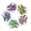
| ||||||||||||||||||||
|---|---|---|---|---|---|---|---|---|---|---|---|---|---|---|---|---|---|---|---|---|---|
| 1 |
| ||||||||||||||||||||
| Unit cell |
| ||||||||||||||||||||
| Noncrystallographic symmetry (NCS) | NCS oper:
|
-Components
| #1: Protein | Mass: 23068.039 Da / Num. of mol.: 5 / Source method: isolated from a natural source / Source: (natural) Homo sapiens (human) / References: UniProt: P02741#2: Chemical | ChemComp-CA /   Mass: 40.078 Da / Num. of mol.: 10 / Source method: obtained synthetically / Formula: Ca Mass: 40.078 Da / Num. of mol.: 10 / Source method: obtained synthetically / Formula: Ca#3: Chemical | ChemComp-PC / 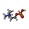  Mass: 184.151 Da / Num. of mol.: 5 / Source method: obtained synthetically / Formula: C5H15NO4P Mass: 184.151 Da / Num. of mol.: 5 / Source method: obtained synthetically / Formula: C5H15NO4PHas protein modification | Y | |
|---|
-Experimental details
-Experiment
| Experiment | Method: X-RAY DIFFRACTION / Number of used crystals: 1 |
|---|
- Sample preparation
| Crystal | Density % sol: 76 % | ||||||||||||||||||||||||||||||||||||
|---|---|---|---|---|---|---|---|---|---|---|---|---|---|---|---|---|---|---|---|---|---|---|---|---|---|---|---|---|---|---|---|---|---|---|---|---|---|
| Crystal grow | pH: 7.6 / Details: pH 7.6 | ||||||||||||||||||||||||||||||||||||
| Crystal grow | *PLUS Method: vapor diffusion, hanging dropDetails: drop is made up of a 1:1 ratio of protein and reservoir solution | ||||||||||||||||||||||||||||||||||||
| Components of the solutions | *PLUS
|
-Data collection
| Diffraction | Mean temperature: 100 K |
|---|---|
| Diffraction source | Source: SYNCHROTRON / Site: SRS  / Beamline: PX9.6 / Wavelength: 0.87 / Beamline: PX9.6 / Wavelength: 0.87 |
| Detector | Type: MARRESEARCH / Detector: IMAGE PLATE / Date: Mar 1, 1995 |
| Radiation | Protocol: SINGLE WAVELENGTH / Monochromatic (M) / Laue (L): M / Scattering type: x-ray |
| Radiation wavelength | Wavelength: 0.87 Å / Relative weight: 1 |
| Reflection | Resolution: 2.5→20 Å / Num. obs: 86545 / % possible obs: 97.6 % / Observed criterion σ(I): 0 / Redundancy: 4.3 % / Rmerge(I) obs: 0.068 / Net I/σ(I): 10.1 |
| Reflection shell | *PLUS % possible obs: 94.5 % |
- Processing
| Software |
| ||||||||||||||||||||||||||||||||||||||||||||||||||||||||||||
|---|---|---|---|---|---|---|---|---|---|---|---|---|---|---|---|---|---|---|---|---|---|---|---|---|---|---|---|---|---|---|---|---|---|---|---|---|---|---|---|---|---|---|---|---|---|---|---|---|---|---|---|---|---|---|---|---|---|---|---|---|---|
| Refinement | Method to determine structure: MOLECULAR REPLACEMENT Starting model: PDB ENTRY 1SAC Resolution: 2.5→20 Å / Cross valid method: THROUGHOUT / σ(F): 3
| ||||||||||||||||||||||||||||||||||||||||||||||||||||||||||||
| Refinement step | Cycle: LAST / Resolution: 2.5→20 Å
| ||||||||||||||||||||||||||||||||||||||||||||||||||||||||||||
| Refine LS restraints |
|
