Movie
Movie Controller

[English] 日本語
 Yorodumi
- EMDB-1876: Structural basis of substrate shuttling in bovine mitochondrial s... -
+ Open data

- Basic information
| Entry | Database: EMDB / ID: EMD-1876 | |||||||||
|---|---|---|---|---|---|---|---|---|---|---|
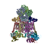
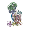
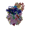
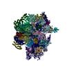
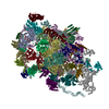
| Title | Structural basis of substrate shuttling in bovine mitochondrial supercomplex I1III2IV1 by single particle cryo-EM | |||||||||
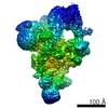
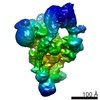
 Map data Map data | 3D cryo-EM map of supercomplex B (I1III2IV1) from bovine heart mitochondria | |||||||||
 Sample Sample |
| |||||||||
 Keywords Keywords | Supercomplex B / mitochondria / respiratory chain / complex I / complex III / complex IV / amphipol A8-35 / random conical tilt | |||||||||
| Function / homology |  Function and homology information Function and homology informationRelease of apoptotic factors from the mitochondria / Formation of apoptosome / Activation of caspases through apoptosome-mediated cleavage / Pyroptosis / Regulation of the apoptosome activity / Transcriptional activation of mitochondrial biogenesis / Detoxification of Reactive Oxygen Species / Complex III assembly / Complex IV assembly / TP53 Regulates Metabolic Genes ...Release of apoptotic factors from the mitochondria / Formation of apoptosome / Activation of caspases through apoptosome-mediated cleavage / Pyroptosis / Regulation of the apoptosome activity / Transcriptional activation of mitochondrial biogenesis / Detoxification of Reactive Oxygen Species / Complex III assembly / Complex IV assembly / TP53 Regulates Metabolic Genes / respiratory chain complex IV assembly / Cytoprotection by HMOX1 / subthalamus development / pons development / NADH dehydrogenase (quinone) (non-electrogenic) activity / mitochondrial respirasome assembly / Translocases; Catalysing the translocation of protons; Linked to oxidoreductase reactions / cerebellar Purkinje cell layer development / Respiratory electron transport / pyramidal neuron development / respiratory chain complex IV / thalamus development / respiratory chain complex / molybdopterin cofactor binding / Mitochondrial translation termination / cytochrome-c oxidase / respiratory chain complex III / oxidative phosphorylation / quinol-cytochrome-c reductase / mitochondrial electron transport, cytochrome c to oxygen / quinol-cytochrome-c reductase activity / cytochrome-c oxidase activity / mitochondrial electron transport, ubiquinol to cytochrome c / iron-sulfur cluster assembly / Mitochondrial protein degradation / hypothalamus development / midbrain development / ubiquinone binding / electron transport coupled proton transport / oxidoreductase activity, acting on NAD(P)H / NADH dehydrogenase activity / respiratory chain complex I / NADH dehydrogenase (ubiquinone) activity / quinone binding / ATP synthesis coupled electron transport / enzyme regulator activity / ferric iron binding / proton transmembrane transport / aerobic respiration / respiratory electron transport chain / central nervous system development / hippocampus development / metalloendopeptidase activity / mitochondrial intermembrane space / 2 iron, 2 sulfur cluster binding / mitochondrial membrane / NAD binding / FMN binding / 4 iron, 4 sulfur cluster binding / electron transfer activity / oxidoreductase activity / mitochondrial inner membrane / iron ion binding / copper ion binding / heme binding / apoptotic process / protein-containing complex / mitochondrion / proteolysis / membrane / metal ion binding / plasma membrane Similarity search - Function | |||||||||
| Biological species |  | |||||||||
| Method | single particle reconstruction / cryo EM / Resolution: 19.0 Å | |||||||||
 Authors Authors | Althoff T / Mills DJ / Popot J-L / Kuehlbrandt W | |||||||||
 Citation Citation | Journal: EMBO J / Year: 2011 Title: Arrangement of electron transport chain components in bovine mitochondrial supercomplex I1III2IV1. Authors: Thorsten Althoff / Deryck J Mills / Jean-Luc Popot / Werner Kühlbrandt /  Abstract: The respiratory chain in the inner mitochondrial membrane contains three large multi-enzyme complexes that together establish the proton gradient for ATP synthesis, and assemble into a supercomplex. ...The respiratory chain in the inner mitochondrial membrane contains three large multi-enzyme complexes that together establish the proton gradient for ATP synthesis, and assemble into a supercomplex. A 19-Å 3D map of the 1.7-MDa amphipol-solubilized supercomplex I(1)III(2)IV(1) from bovine heart obtained by single-particle electron cryo-microscopy reveals an amphipol belt replacing the membrane lipid bilayer. A precise fit of the X-ray structures of complex I, the complex III dimer, and monomeric complex IV indicates distances of 13 nm between the ubiquinol-binding sites of complexes I and III, and of 10-11 nm between the cytochrome c binding sites of complexes III and IV. The arrangement of respiratory chain complexes suggests two possible pathways for efficient electron transfer through the supercomplex, of which the shorter branch through the complex III monomer proximal to complex I may be preferred. #1: Year: 2011Title: Strukturelle Untersuchungen am Superkomplex I1III2IV1 der Atmungskette mittels Kryoelektronenmikroskopie Authors: Althoff T | |||||||||
| History |
|
- Structure visualization
| Movie |
Movie viewer |
|---|---|
| Structure viewer | EM map: SurfViewMolmilJmol/JSmol |
| Supplemental images |
 UCSF Chimera
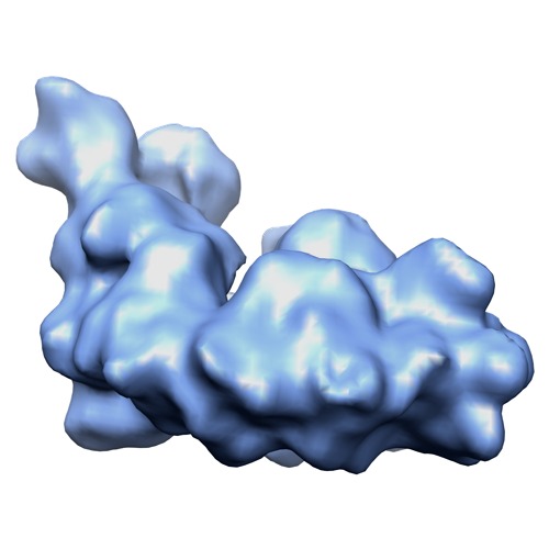
- Downloads & links
-EMDB archive
| Map data | emd_1876.map.gz | 250.7 KB | EMDB map data format | |
|---|---|---|---|---|
| Header (meta data) | emd-1876-v30.xmlemd-1876.xml | 20.2 KB 20.2 KB | Display Display | EMDB header |
| Images | EMD1876_bt_mito_supercomplex-b_cryo_front-view.tif | 160.4 KB | ||
| Archive directory |  http://ftp.pdbj.org/pub/emdb/structures/EMD-1876ftp://ftp.pdbj.org/pub/emdb/structures/EMD-1876 http://ftp.pdbj.org/pub/emdb/structures/EMD-1876ftp://ftp.pdbj.org/pub/emdb/structures/EMD-1876 | HTTPS FTP |
-Related structure data
| Related structure data |  2ybbMC M: atomic model generated by this map C: citing same article ( |
|---|---|
| Similar structure data |

-Links
| EMDB pages | EMDB (EBI/PDBe) / EMDataResource |
|---|---|
| Related items in Molecule of the Month |


-Map
| File | Download / File: emd_1876.map.gz / Format: CCP4 / Size: 5.2 MB / Type: IMAGE STORED AS FLOATING POINT NUMBER (4 BYTES) | ||||||||||||||||||||||||||||||||||||||||||||||||||||||||||||||||||||
|---|---|---|---|---|---|---|---|---|---|---|---|---|---|---|---|---|---|---|---|---|---|---|---|---|---|---|---|---|---|---|---|---|---|---|---|---|---|---|---|---|---|---|---|---|---|---|---|---|---|---|---|---|---|---|---|---|---|---|---|---|---|---|---|---|---|---|---|---|---|
| Annotation | 3D cryo-EM map of supercomplex B (I1III2IV1) from bovine heart mitochondria | ||||||||||||||||||||||||||||||||||||||||||||||||||||||||||||||||||||
| Projections & slices | Image control
Images are generated by Spider. | ||||||||||||||||||||||||||||||||||||||||||||||||||||||||||||||||||||
| Voxel size | X=Y=Z: 4.76 Å | ||||||||||||||||||||||||||||||||||||||||||||||||||||||||||||||||||||
| Density |
| ||||||||||||||||||||||||||||||||||||||||||||||||||||||||||||||||||||
| Symmetry | Space group: 1 | ||||||||||||||||||||||||||||||||||||||||||||||||||||||||||||||||||||
| Details | EMDB XML:
CCP4 map header:
| ||||||||||||||||||||||||||||||||||||||||||||||||||||||||||||||||||||
 Z (Sec.)
 X (Row.)
 Y (Col.)


-Supplemental data
- Sample components
-Entire : Respiratory supercomplex B (I1III2IV1) composed of complex I, dim...
| Entire | Name: Respiratory supercomplex B (I1III2IV1) composed of complex I, dimeric complex III and complex IV from bovine heart mitochondria |
|---|---|
| Components |
|
-Supramolecule #1000: Respiratory supercomplex B (I1III2IV1) composed of complex I, dim...
| Supramolecule | Name: Respiratory supercomplex B (I1III2IV1) composed of complex I, dimeric complex III and complex IV from bovine heart mitochondria type: sample / ID: 1000 Details: The supercomplexes were kept soluble by amphipol A8-35 Oligomeric state: A monomer of complex I assembles with a dimer of complex III and a monomer of complex IV Number unique components: 4 |
|---|---|
| Molecular weight | Theoretical: 1.7 MDa |
-Macromolecule #1: NADH-dehydrogenase
| Macromolecule | Name: NADH-dehydrogenase / type: protein_or_peptide / ID: 1 / Name.synonym: Complex I / Number of copies: 1 / Oligomeric state: Monomer / Recombinant expression: No |
|---|---|
| Source (natural) | Organism: |
| Molecular weight | Experimental: 980 KDa / Theoretical: 980 KDa |
-Macromolecule #2: Cytochrome c reductase
| Macromolecule | Name: Cytochrome c reductase / type: protein_or_peptide / ID: 2 / Name.synonym: Complex III / Number of copies: 2 / Oligomeric state: Dimer / Recombinant expression: No |
|---|---|
| Source (natural) | Organism: |
| Molecular weight | Experimental: 243 KDa / Theoretical: 241 KDa |
-Macromolecule #3: Cytochrome c oxidase
| Macromolecule | Name: Cytochrome c oxidase / type: protein_or_peptide / ID: 3 / Name.synonym: Complex IV / Number of copies: 1 / Oligomeric state: Monomer / Recombinant expression: No |
|---|---|
| Source (natural) | Organism: |
| Molecular weight | Experimental: 207 KDa / Theoretical: 204 KDa |
-Macromolecule #4: Cytochrome c
| Macromolecule | Name: Cytochrome c / type: protein_or_peptide / ID: 4 / Name.synonym: Cytochrome c / Number of copies: 1 / Oligomeric state: Monomer / Recombinant expression: No |
|---|---|
| Source (natural) | Organism: |
| Molecular weight | Experimental: 12 KDa / Theoretical: 12 KDa |
 Processing
 Electron microscopy