Movie
Movie Controller


+ Open data

- Basic information
| Entry | Database: EMDB / ID: EMD-1764 | |||||||||
|---|---|---|---|---|---|---|---|---|---|---|
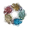
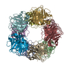
| Title | Single particle analysis of Kir2.1NC_4 in negative stain | |||||||||
 Map data Map data | 3d map of the Kir2.1NC_4 tetramer, C_4 symmetry applied | |||||||||
 Sample Sample |
| |||||||||
 Keywords Keywords | Ion Channel / cytoplasmic domain / inwardly rectifying / membrane protein / homotetramer | |||||||||
| Function / homology |  Function and homology information Function and homology informationClassical Kir channels / regulation of skeletal muscle contraction via regulation of action potential / relaxation of skeletal muscle / Phase 4 - resting membrane potential / cardiac muscle cell action potential / voltage-gated potassium channel activity involved in cardiac muscle cell action potential repolarization / Activation of G protein gated Potassium channels / Inhibition of voltage gated Ca2+ channels via Gbeta/gamma subunits / membrane repolarization during action potential / magnesium ion transport ...Classical Kir channels / regulation of skeletal muscle contraction via regulation of action potential / relaxation of skeletal muscle / Phase 4 - resting membrane potential / cardiac muscle cell action potential / voltage-gated potassium channel activity involved in cardiac muscle cell action potential repolarization / Activation of G protein gated Potassium channels / Inhibition of voltage gated Ca2+ channels via Gbeta/gamma subunits / membrane repolarization during action potential / magnesium ion transport / membrane repolarization during cardiac muscle cell action potential / regulation of membrane repolarization / inward rectifier potassium channel activity / positive regulation of potassium ion transmembrane transport / cardiac muscle cell action potential involved in contraction / regulation of cardiac muscle cell contraction / relaxation of cardiac muscle / potassium ion import across plasma membrane / regulation of heart rate by cardiac conduction / intercalated disc / membrane => GO:0016020 / phosphatidylinositol-4,5-bisphosphate binding / voltage-gated potassium channel complex / potassium ion transmembrane transport / T-tubule / potassium ion transport / cellular response to mechanical stimulus / protein homotetramerization / dendritic spine / postsynaptic membrane / neuronal cell body / dendrite / glutamatergic synapse / membrane / identical protein binding / plasma membrane Similarity search - Function | |||||||||
| Biological species |  | |||||||||
| Method | single particle reconstruction / negative staining / Resolution: 17.2 Å | |||||||||
 Authors Authors | Fomina S / Howard TD / Sleator OK / Golovanova M / O'Ryan L / Leyland M / Grossmann JG / Collins RF / Prince SM | |||||||||
 Citation Citation | Journal: Biochim Biophys Acta / Year: 2011 Title: Self-directed assembly and clustering of the cytoplasmic domains of inwardly rectifying Kir2.1 potassium channels on association with PSD-95. Authors: Svetlana Fomina / Tina D Howard / Olivia K Sleator / Marina Golovanova / Liam O'Ryan / Mark L Leyland / J Günter Grossmann / Richard F Collins / Stephen M Prince /  Abstract: The interaction of the extra-membranous domain of tetrameric inwardly rectifying Kir2.1 ion channels (Kir2.1NC(4)) with the membrane associated guanylate kinase protein PSD-95 has been studied using ...The interaction of the extra-membranous domain of tetrameric inwardly rectifying Kir2.1 ion channels (Kir2.1NC(4)) with the membrane associated guanylate kinase protein PSD-95 has been studied using Transmission Electron Microscopy in negative stain. Three types of complexes were observed in electron micrographs corresponding to a 1:1 complex, a large self-enclosed tetrad complex and extended chains of linked channel domains. Using models derived from small angle X-ray scattering experiments in which high resolution structures from X-ray crystallographic and Nuclear Magnetic Resonance studies are positioned, the envelopes from single particle analysis can be resolved as a Kir2.1NC(4):PSD-95 complex and a tetrad of this unit (Kir2.1NC(4):PSD-95)(4). The tetrad complex shows the close association of the Kir2.1 cytoplasmic domains and the influence of PSD-95 mediated self-assembly on the clustering of these channels. | |||||||||
| History |
|
- Structure visualization
| Movie |
Movie viewer |
|---|---|
| Structure viewer | EM map: SurfViewMolmilJmol/JSmol |
| Supplemental images |
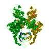
 UCSF Chimera
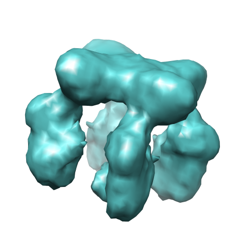
- Downloads & links
-EMDB archive
| Map data | emd_1764.map.gz | 508.6 KB | EMDB map data format | |
|---|---|---|---|---|
| Header (meta data) | emd-1764-v30.xmlemd-1764.xml | 10.7 KB 10.7 KB | Display Display | EMDB header |
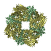
| Images |  emd_1764.png emd_1764.png | 105.4 KB | ||
| Archive directory |  http://ftp.pdbj.org/pub/emdb/structures/EMD-1764ftp://ftp.pdbj.org/pub/emdb/structures/EMD-1764 http://ftp.pdbj.org/pub/emdb/structures/EMD-1764ftp://ftp.pdbj.org/pub/emdb/structures/EMD-1764 | HTTPS FTP |
-Related structure data
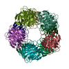
| Related structure data |  2xkyMC 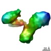 1761C 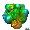 1765C 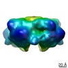 1766C 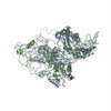 2xkxC M: atomic model generated by this map C: citing same article ( |
|---|---|
| Similar structure data |

-Links
| EMDB pages | EMDB (EBI/PDBe) / EMDataResource |
|---|---|
| Related items in Molecule of the Month |

-Map
| File | Download / File: emd_1764.map.gz / Format: CCP4 / Size: 1001 KB / Type: IMAGE STORED AS FLOATING POINT NUMBER (4 BYTES) | ||||||||||||||||||||||||||||||||||||||||||||||||||||||||||||||||||||
|---|---|---|---|---|---|---|---|---|---|---|---|---|---|---|---|---|---|---|---|---|---|---|---|---|---|---|---|---|---|---|---|---|---|---|---|---|---|---|---|---|---|---|---|---|---|---|---|---|---|---|---|---|---|---|---|---|---|---|---|---|---|---|---|---|---|---|---|---|---|
| Annotation | 3d map of the Kir2.1NC_4 tetramer, C_4 symmetry applied | ||||||||||||||||||||||||||||||||||||||||||||||||||||||||||||||||||||
| Projections & slices | Image control
Images are generated by Spider. | ||||||||||||||||||||||||||||||||||||||||||||||||||||||||||||||||||||
| Voxel size | X=Y=Z: 2.93 Å | ||||||||||||||||||||||||||||||||||||||||||||||||||||||||||||||||||||
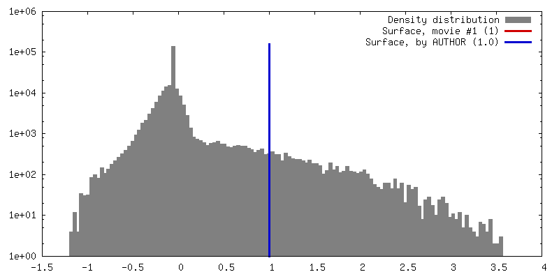
| Density |
| ||||||||||||||||||||||||||||||||||||||||||||||||||||||||||||||||||||
| Symmetry | Space group: 1 | ||||||||||||||||||||||||||||||||||||||||||||||||||||||||||||||||||||
| Details | EMDB XML:
CCP4 map header:
| ||||||||||||||||||||||||||||||||||||||||||||||||||||||||||||||||||||
 Z (Sec.)
 Y (Row.)
 X (Col.)


-Supplemental data
- Sample components
-Entire : mouse Kir2.1, cytoplamic domain, homotetramer of fused N,C termini
| Entire | Name: mouse Kir2.1, cytoplamic domain, homotetramer of fused N,C termini |
|---|---|
| Components |
|
-Supramolecule #1000: mouse Kir2.1, cytoplamic domain, homotetramer of fused N,C termini
| Supramolecule | Name: mouse Kir2.1, cytoplamic domain, homotetramer of fused N,C termini type: sample / ID: 1000 / Oligomeric state: Tetramer / Number unique components: 1 |
|---|---|
| Molecular weight | Experimental: 140 KDa / Theoretical: 140 KDa / Method: SDS-PAGE for Kir2.1NC construct x 4 |
-Macromolecule #1: Kir2.1 cytoplasmic domain
| Macromolecule | Name: Kir2.1 cytoplasmic domain / type: protein_or_peptide / ID: 1 / Name.synonym: Kir2.1NC / Number of copies: 4 / Oligomeric state: Tetramer / Recombinant expression: Yes |
|---|---|
| Source (natural) | Organism: |
| Molecular weight | Experimental: 140 KDa / Theoretical: 140 KDa |
| Recombinant expression | Organism:  |
| Sequence | GO: membrane => GO:0016020 / InterPro: Potassium channel, inwardly rectifying, Kir2.1 |
-Experimental details
-Structure determination
| Method | negative staining |
|---|---|
 Processing Processing | single particle reconstruction |
| Aggregation state | particle |
-Sample preparation
| Concentration | 1 mg/mL |
|---|---|
| Buffer | pH: 7.5 Details: 20mM Tris/HCl, 150mM NaCl, 1mM reduced GSH, 1mM EDTA, 50mM L-Glutamic acid, 50mM L-Arginine |
| Staining | Type: NEGATIVE Details: Samples were adsorbed onto freshly glow discharged carbon film grids. Sample solution was pipetted into the grid followed by blotting, de-ionized water was then applied for 10s followed by ...Details: Samples were adsorbed onto freshly glow discharged carbon film grids. Sample solution was pipetted into the grid followed by blotting, de-ionized water was then applied for 10s followed by blotting, 2% w/v Uranyl Acetate solution was applied followed by a final blotting step. |
| Grid | Details: 400 mesh Copper |
| Vitrification | Cryogen name: NONE / Instrument: OTHER |
- Electron microscopy
| Microscope | FEI TECNAI 10 |
|---|---|
| Details | Low dose |
| Image recording | Category: FILM / Film or detector model: GATAN ORIUS SC200 (2k x 2k) / Digitization - Scanner: OTHER / Digitization - Sampling interval: 10 µm / Number real images: 22 / Bits/pixel: 8 |
| Tilt angle min | 0 |
| Tilt angle max | 0 |
| Electron beam | Acceleration voltage: 100 kV / Electron source: TUNGSTEN HAIRPIN |
| Electron optics | Illumination mode: FLOOD BEAM / Imaging mode: BRIGHT FIELD / Cs: 2.0 mm / Nominal defocus max: 1.25 µm / Nominal defocus min: 0.6 µm |
| Sample stage | Specimen holder: Eucentric / Specimen holder model: JEOL |
-Image processing
| Details | Particles initially selected using automated particle picking based on a subset of representative particles on a single micrograph, followed by model-based picking. |
|---|---|
| CTF correction | Details: Parameters determined using Scattering curve |
| Final reconstruction | Applied symmetry - Point group: C4 (4 fold cyclic) / Resolution.type: BY AUTHOR / Resolution: 17.2 Å / Resolution method: FSC 0.5 CUT-OFF / Software - Name: EMAN / Number images used: 49012 |
| Final two d classification | Number classes: 62 |