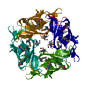
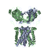
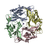
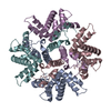
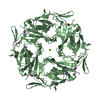
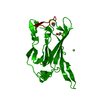
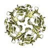
Journal: Nat Neurosci / Year: 2005 Title: Cytoplasmic domain structures of Kir2.1 and Kir3.1 show sites for modulating gating and rectification. Authors: Scott Pegan / Christine Arrabit / Wei Zhou / Witek Kwiatkowski / Anthony Collins / Paul A Slesinger / Senyon Choe / Abstract: N- and C-terminal cytoplasmic domains of inwardly rectifying K (Kir) channels control the ion-permeation pathway through diverse interactions with small molecules and protein ligands in the cytoplasm. ...N- and C-terminal cytoplasmic domains of inwardly rectifying K (Kir) channels control the ion-permeation pathway through diverse interactions with small molecules and protein ligands in the cytoplasm. Two new crystal structures of the cytoplasmic domains of Kir2.1 (Kir2.1(L)) and the G protein-sensitive Kir3.1 (Kir3.1(S)) channels in the absence of PIP(2) show the cytoplasmic ion-permeation pathways occluded by four cytoplasmic loops that form a girdle around the central pore (G-loop). Significant flexibility of the pore-facing G-loop of Kir2.1(L) and Kir3.1(S) suggests a possible role as a diffusion barrier between cytoplasmic and transmembrane pores. Consistent with this, mutations of the G-loop disrupted gating or inward rectification. Structural comparison shows a di-aspartate cluster on the distal end of the cytoplasmic pore of Kir2.1(L) that is important for modulating inward rectification. Taken together, these results suggest the cytoplasmic domains of Kir channels undergo structural changes to modulate gating and inward rectification.
In the structure databanks used in Yorodumi, some data are registered as the other names, "COVID-19 virus" and "2019-nCoV". Here are the details of the virus and the list of structure data.
Jan 31, 2019. EMDB accession codes are about to change! (news from PDBe EMDB page)
EMDB accession codes are about to change! (news from PDBe EMDB page)
The allocation of 4 digits for EMDB accession codes will soon come to an end. Whilst these codes will remain in use, new EMDB accession codes will include an additional digit and will expand incrementally as the available range of codes is exhausted. The current 4-digit format prefixed with “EMD-” (i.e. EMD-XXXX) will advance to a 5-digit format (i.e. EMD-XXXXX), and so on. It is currently estimated that the 4-digit codes will be depleted around Spring 2019, at which point the 5-digit format will come into force.
The EM Navigator/Yorodumi systems omit the EMD- prefix.
Related info.:Q: What is EMD? / ID/Accession-code notation in Yorodumi/EM Navigator
Yorodumi is a browser for structure data from EMDB, PDB, SASBDB, etc.
This page is also the successor to EM Navigator detail page, and also detail information page/front-end page for Omokage search.
The word "yorodu" (or yorozu) is an old Japanese word meaning "ten thousand". "mi" (miru) is to see.
Related info.:EMDB / PDB / SASBDB / Comparison of 3 databanks / Yorodumi Search / Aug 31, 2016. New EM Navigator & Yorodumi / Yorodumi Papers / Jmol/JSmol / Function and homology information / Changes in new EM Navigator and Yorodumi
 Movie
Movie Controller

 Yorodumi
 Open data
 Basic information
 Components
 Keywords
 Function and homology information
 X-RAY DIFFRACTION /
  Authors
 Citation
 Structure visualization
 Downloads & links
 Other downloads
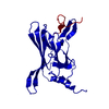


 PDBj
PDBj
 Assembly
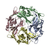


 Mass: 18.015 Da / Num. of mol.: 213 / Source method: isolated from a natural source / Formula: H2O
 Sample preparation
 Processing