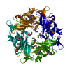
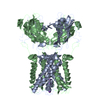
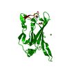
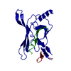
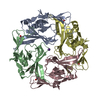
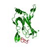
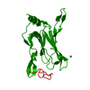
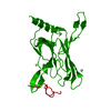
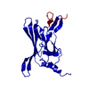
Journal: Nat Neurosci / Year: 2005 Title: Cytoplasmic domain structures of Kir2.1 and Kir3.1 show sites for modulating gating and rectification. Authors: Scott Pegan / Christine Arrabit / Wei Zhou / Witek Kwiatkowski / Anthony Collins / Paul A Slesinger / Senyon Choe / Abstract: N- and C-terminal cytoplasmic domains of inwardly rectifying K (Kir) channels control the ion-permeation pathway through diverse interactions with small molecules and protein ligands in the cytoplasm. ...N- and C-terminal cytoplasmic domains of inwardly rectifying K (Kir) channels control the ion-permeation pathway through diverse interactions with small molecules and protein ligands in the cytoplasm. Two new crystal structures of the cytoplasmic domains of Kir2.1 (Kir2.1(L)) and the G protein-sensitive Kir3.1 (Kir3.1(S)) channels in the absence of PIP(2) show the cytoplasmic ion-permeation pathways occluded by four cytoplasmic loops that form a girdle around the central pore (G-loop). Significant flexibility of the pore-facing G-loop of Kir2.1(L) and Kir3.1(S) suggests a possible role as a diffusion barrier between cytoplasmic and transmembrane pores. Consistent with this, mutations of the G-loop disrupted gating or inward rectification. Structural comparison shows a di-aspartate cluster on the distal end of the cytoplasmic pore of Kir2.1(L) that is important for modulating inward rectification. Taken together, these results suggest the cytoplasmic domains of Kir channels undergo structural changes to modulate gating and inward rectification.
History
Deposition
Jul 24, 2004
Deposition site: RCSB / Processing site: RCSB
Revision 1.0
Mar 8, 2005
Provider: repository / Type: Initial release
Revision 1.1
Apr 30, 2008
Group: Version format compliance
Revision 1.2
Jul 13, 2011
Group: Derived calculations / Version format compliance
Revision 1.3
Aug 23, 2017
Group: Source and taxonomy / Category: entity_src_gen
Resolution: 2.09→91.29 Å / Cor.coef. Fo:Fc: 0.95 / Cor.coef. Fo:Fc free: 0.936 / SU B: 4.589 / SU ML: 0.121 / Cross valid method: THROUGHOUT / ESU R: 0.191 / ESU R Free: 0.171 / Stereochemistry target values: MAXIMUM LIKELIHOOD / Details: HYDROGENS HAVE BEEN ADDED IN THE RIDING POSITIONS
Rfactor
Num. reflection
% reflection
Selection details
Rfree
0.23505
813
5.1 %
RANDOM
Rwork
0.19431
-
-
-
all
0.19641
15270
-
-
obs
0.19641
15270
96 %
-
Solvent computation
Ion probe radii: 0.8 Å / Shrinkage radii: 0.8 Å / VDW probe radii: 1.4 Å / Solvent model: BABINET MODEL WITH MASK
Displacement parameters
Biso mean: 33.928 Å2
Baniso -1
Baniso -2
Baniso -3
1-
-1.12 Å2
0 Å2
0 Å2
2-
-
-1.12 Å2
0 Å2
3-
-
-
2.23 Å2
Refinement step
Cycle: LAST / Resolution: 2.09→91.29 Å
Protein
Nucleic acid
Ligand
Solvent
Total
Num. atoms
1599
0
0
129
1728
Refine LS restraints
Refine-ID
Type
Dev ideal
Dev ideal target
Number
X-RAY DIFFRACTION
r_bond_refined_d
0.034
0.021
1632
X-RAY DIFFRACTION
r_bond_other_d
0.002
0.02
1461
X-RAY DIFFRACTION
r_angle_refined_deg
2.378
1.951
2204
X-RAY DIFFRACTION
r_angle_other_deg
1.161
3
3398
X-RAY DIFFRACTION
r_dihedral_angle_1_deg
9.008
5
198
X-RAY DIFFRACTION
r_dihedral_angle_2_deg
X-RAY DIFFRACTION
r_dihedral_angle_3_deg
X-RAY DIFFRACTION
r_dihedral_angle_4_deg
X-RAY DIFFRACTION
r_chiral_restr
0.177
0.2
246
X-RAY DIFFRACTION
r_gen_planes_refined
0.012
0.02
1803
X-RAY DIFFRACTION
r_gen_planes_other
0.005
0.02
340
X-RAY DIFFRACTION
r_nbd_refined
0.234
0.2
305
X-RAY DIFFRACTION
r_nbd_other
0.272
0.2
1720
X-RAY DIFFRACTION
r_nbtor_refined
X-RAY DIFFRACTION
r_nbtor_other
0.098
0.2
992
X-RAY DIFFRACTION
r_xyhbond_nbd_refined
0.222
0.2
112
X-RAY DIFFRACTION
r_xyhbond_nbd_other
X-RAY DIFFRACTION
r_metal_ion_refined
X-RAY DIFFRACTION
r_metal_ion_other
X-RAY DIFFRACTION
r_symmetry_vdw_refined
0.237
0.2
14
X-RAY DIFFRACTION
r_symmetry_vdw_other
0.377
0.2
79
X-RAY DIFFRACTION
r_symmetry_hbond_refined
0.2
0.2
21
X-RAY DIFFRACTION
r_symmetry_hbond_other
X-RAY DIFFRACTION
r_mcbond_it
1.858
1.5
996
X-RAY DIFFRACTION
r_mcbond_other
X-RAY DIFFRACTION
r_mcangle_it
3.268
2
1618
X-RAY DIFFRACTION
r_scbond_it
4.212
3
636
X-RAY DIFFRACTION
r_scangle_it
6.655
4.5
586
X-RAY DIFFRACTION
r_rigid_bond_restr
X-RAY DIFFRACTION
r_sphericity_free
X-RAY DIFFRACTION
r_sphericity_bonded
LS refinement shell
Resolution: 2.09→2.145 Å / Total num. of bins used: 20 /
Rfactor
Num. reflection
Rfree
0.322
47
Rwork
0.276
999
+
About Yorodumi
-
News
-
Feb 9, 2022. New format data for meta-information of EMDB entries
New format data for meta-information of EMDB entries
Version 3 of the EMDB header file is now the official format.
The previous official version 1.9 will be removed from the archive.
In the structure databanks used in Yorodumi, some data are registered as the other names, "COVID-19 virus" and "2019-nCoV". Here are the details of the virus and the list of structure data.
Jan 31, 2019. EMDB accession codes are about to change! (news from PDBe EMDB page)
EMDB accession codes are about to change! (news from PDBe EMDB page)
The allocation of 4 digits for EMDB accession codes will soon come to an end. Whilst these codes will remain in use, new EMDB accession codes will include an additional digit and will expand incrementally as the available range of codes is exhausted. The current 4-digit format prefixed with “EMD-” (i.e. EMD-XXXX) will advance to a 5-digit format (i.e. EMD-XXXXX), and so on. It is currently estimated that the 4-digit codes will be depleted around Spring 2019, at which point the 5-digit format will come into force.
The EM Navigator/Yorodumi systems omit the EMD- prefix.
Related info.:Q: What is EMD? / ID/Accession-code notation in Yorodumi/EM Navigator
Yorodumi is a browser for structure data from EMDB, PDB, SASBDB, etc.
This page is also the successor to EM Navigator detail page, and also detail information page/front-end page for Omokage search.
The word "yorodu" (or yorozu) is an old Japanese word meaning "ten thousand". "mi" (miru) is to see.
Related info.:EMDB / PDB / SASBDB / Comparison of 3 databanks / Yorodumi Search / Aug 31, 2016. New EM Navigator & Yorodumi / Yorodumi Papers / Jmol/JSmol / Function and homology information / Changes in new EM Navigator and Yorodumi
 Movie
Movie Controller


 Open data
 Basic information
 Components
 Keywords
 Function and homology information
 X-RAY DIFFRACTION /
  Authors
 Citation
 Structure visualization
 Downloads & links
 Other downloads
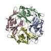


 PDBj
PDBj
 Assembly

 Mass: 18.015 Da / Num. of mol.: 129 / Source method: isolated from a natural source / Formula: H2O
 Sample preparation
 Processing