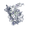
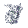
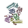
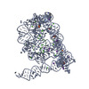
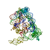
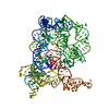
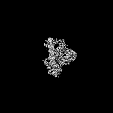
Journal: J Virol / Year: 2021 Title: Structures of Substrate Complexes of Foamy Viral Protease-Reverse Transcriptase. Authors: Marzena Nowacka / Elżbieta Nowak / Mariusz Czarnocki-Cieciura / Justyna Jackiewicz / Krzysztof Skowronek / Roman H Szczepanowski / Birgitta M Wöhrl / Marcin Nowotny / Abstract: Reverse transcriptases (RTs) use their DNA polymerase and RNase H activities to catalyze the conversion of single-stranded RNA to double-stranded DNA (dsDNA), a crucial process for the replication of ...Reverse transcriptases (RTs) use their DNA polymerase and RNase H activities to catalyze the conversion of single-stranded RNA to double-stranded DNA (dsDNA), a crucial process for the replication of retroviruses. Foamy viruses (FVs) possess a unique RT, which is a fusion with the protease (PR) domain. The mechanism of substrate binding by this enzyme has been unknown. Here, we report a crystal structure of monomeric full-length marmoset FV (MFV) PR-RT in complex with an RNA/DNA hybrid substrate. We also describe a structure of MFV PR-RT with an RNase H deletion in complex with a dsDNA substrate in which the enzyme forms an asymmetric homodimer. Cryo-electron microscopy reconstruction of the full-length MFV PR-RT-dsDNA complex confirmed the dimeric architecture. These findings represent the first structural description of nucleic acid binding by a foamy viral RT and demonstrate its ability to change its oligomeric state depending on the type of bound nucleic acid. Reverse transcriptases (RTs) are intriguing enzymes converting single-stranded RNA to dsDNA. Their activity is essential for retroviruses, which are divided into two subfamilies differing significantly in their life cycles: and . The latter family is much more ancient and comprises five genera. A unique feature of foamy viral RTs is that they contain N-terminal protease (PR) domains, which are not present in orthoretroviral enzymes. So far, no structural information for full-length foamy viral PR-RT interacting with nucleic substrates has been reported. Here, we present crystal and cryo-electron microscopy structures of marmoset foamy virus (MFV) PR-RT. These structures revealed the mode of binding of RNA/DNA and dsDNA substrates. Moreover, unexpectedly, the structures and biochemical data showed that foamy viral PR-RT can adopt both a monomeric configuration, which is observed in our structures in the presence of an RNA/DNA hybrid, and an asymmetric dimer arrangement, which we observed in the presence of dsDNA.
History
Deposition
Mar 30, 2021
-
Header (metadata) release
Jun 30, 2021
-
Map release
Jun 30, 2021
-
Update
Jul 9, 2025
-
Current status
Jul 9, 2025
Processing site: PDBe / Status: Released
-
Structure visualization
Movie
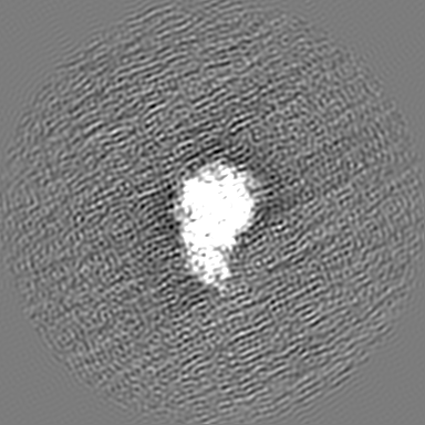
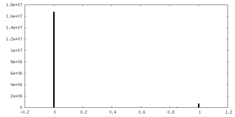
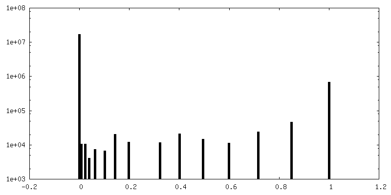
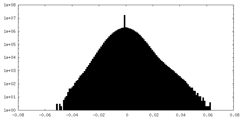
Surface view with section colored by density value
Macromolecule #2: DNA (5'-D(*AP*AP*CP*AP*GP*AP*GP*TP*GP*CP*GP*AP*CP*AP*CP*CP*TP*GP*...
Macromolecule
Name: DNA (5'-D(*AP*AP*CP*AP*GP*AP*GP*TP*GP*CP*GP*AP*CP*AP*CP*CP*TP*GP*AP*TP*TP*CP*CP*A)-3') type: dna / ID: 2 / Number of copies: 1 / Classification: DNA
In the structure databanks used in Yorodumi, some data are registered as the other names, "COVID-19 virus" and "2019-nCoV". Here are the details of the virus and the list of structure data.
Jan 31, 2019. EMDB accession codes are about to change! (news from PDBe EMDB page)
EMDB accession codes are about to change! (news from PDBe EMDB page)
The allocation of 4 digits for EMDB accession codes will soon come to an end. Whilst these codes will remain in use, new EMDB accession codes will include an additional digit and will expand incrementally as the available range of codes is exhausted. The current 4-digit format prefixed with “EMD-” (i.e. EMD-XXXX) will advance to a 5-digit format (i.e. EMD-XXXXX), and so on. It is currently estimated that the 4-digit codes will be depleted around Spring 2019, at which point the 5-digit format will come into force.
The EM Navigator/Yorodumi systems omit the EMD- prefix.
Related info.:Q: What is EMD? / ID/Accession-code notation in Yorodumi/EM Navigator
Yorodumi is a browser for structure data from EMDB, PDB, SASBDB, etc.
This page is also the successor to EM Navigator detail page, and also detail information page/front-end page for Omokage search.
The word "yorodu" (or yorozu) is an old Japanese word meaning "ten thousand". "mi" (miru) is to see.
Related info.:EMDB / PDB / SASBDB / Comparison of 3 databanks / Yorodumi Search / Aug 31, 2016. New EM Navigator & Yorodumi / Yorodumi Papers / Jmol/JSmol / Function and homology information / Changes in new EM Navigator and Yorodumi
 Movie
Movie Controller

 Yorodumi
 Open data
 Basic information
 Map data
 Sample
 Keywords
 Function and homology information
 White-tufted-ear marmoset simian foamy virus / synthetic construct (others)
 Authors
 Poland, 1 items
  Citation
 Structure visualization
 Downloads & links
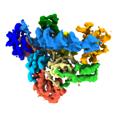 emd_12698.png
 http://ftp.pdbj.org/pub/emdb/structures/EMD-12698


 Z (Sec.)
 Y (Row.)
 X (Col.)


 Sample components
 Processing
 Electron microscopy
 FIELD EMISSION GUN