 Movie
Movie Controller

[English] 日本語
 Yorodumi
- PDB-4e8v: Structure of Oceanobacillus iheyensis group II intron in a ligand... -
+ Open data

- Basic information
| Entry | Database: PDB / ID: 4e8v | ||||||
|---|---|---|---|---|---|---|---|
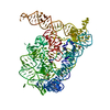
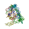
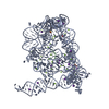
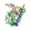
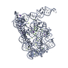
| Title | Structure of Oceanobacillus iheyensis group II intron in a ligand-free state in the presence of K+ and Ba2+ | ||||||
 Components Components | Group IIC intron | ||||||
 Keywords Keywords | RNA / ribozyme / metalloenzyme / self-splicing / retrotransposition | ||||||
| Function / homology | : / : / RNA / RNA (> 10) / RNA (> 100) Function and homology information Function and homology information | ||||||
| Biological species |  Oceanobacillus iheyensis (bacteria) Oceanobacillus iheyensis (bacteria) | ||||||
| Method |  X-RAY DIFFRACTION / SYNCHROTRON / MOLECULAR REPLACEMENT / Resolution: 3.995 Å X-RAY DIFFRACTION / SYNCHROTRON / MOLECULAR REPLACEMENT / Resolution: 3.995 Å | ||||||
 Authors Authors | Marcia, M. / Pyle, A.M. | ||||||
 Citation Citation | Journal: Cell(Cambridge,Mass.) / Year: 2012 Title: Visualizing Group II Intron Catalysis through the Stages of Splicing. Authors: Marcia, M. / Pyle, A.M. | ||||||
| History |
|
- Structure visualization
| Structure viewer | Molecule: MolmilJmol/JSmol |
|---|
- Downloads & links
-Download
| PDBx/mmCIF format | 4e8v.cif.gz | 228.7 KB | Display | PDBx/mmCIF format |
|---|---|---|---|---|
| PDB format | pdb4e8v.ent.gz | 174.8 KB | Display | PDB format |
| PDBx/mmJSON format | 4e8v.json.gz | Tree view | PDBx/mmJSON format | |
| Others |  Other downloads Other downloads |
-Validation report
| Arichive directory | https://data.pdbj.org/pub/pdb/validation_reports/e8/4e8vftp://data.pdbj.org/pub/pdb/validation_reports/e8/4e8v | HTTPS FTP |
|---|
-Related structure data
| Related structure data | 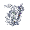 4e8kC 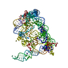 4e8mC 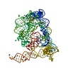 4e8nC  4e8pC  4e8qC  4e8rC  4e8tC 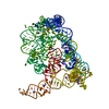 4faqC  4farC  4fauC  4fawC  4faxC 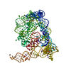 4fb0C  3igiS C: citing same article ( S: Starting model for refinement |
|---|---|
| Similar structure data |


-Links
 PDBj
PDBj


- Assembly
| Deposited unit | 
| ||||||||
|---|---|---|---|---|---|---|---|---|---|
| 1 |
| ||||||||
| Unit cell |
|
-Components
-RNA chain , 1 types, 1 molecules A
| #1: RNA chain | Mass: 127646.586 Da / Num. of mol.: 1 / Fragment: domains 1-5 / Source method: obtained synthetically / Details: in vitro transcription / Source: (synth.) Oceanobacillus iheyensis (bacteria) |
|---|
-Non-polymers , 5 types, 86 molecules 


| #2: Chemical | ChemComp-MG /  Mass: 24.305 Da / Num. of mol.: 5 / Source method: obtained synthetically / Formula: Mg Mass: 24.305 Da / Num. of mol.: 5 / Source method: obtained synthetically / Formula: Mg#3: Chemical | ChemComp-BA /  Mass: 137.327 Da / Num. of mol.: 42 / Source method: obtained synthetically / Formula: Ba Mass: 137.327 Da / Num. of mol.: 42 / Source method: obtained synthetically / Formula: Ba#4: Chemical | ChemComp-K /  Mass: 39.098 Da / Num. of mol.: 4 / Source method: obtained synthetically / Formula: K Mass: 39.098 Da / Num. of mol.: 4 / Source method: obtained synthetically / Formula: K#5: Chemical | ChemComp-EPE /  Mass: 238.305 Da / Num. of mol.: 6 / Source method: obtained synthetically / Formula: C8H18N2O4S / Comment: pH buffer*YM Mass: 238.305 Da / Num. of mol.: 6 / Source method: obtained synthetically / Formula: C8H18N2O4S / Comment: pH buffer*YM#6: Water | ChemComp-HOH / | Mass: 18.015 Da / Num. of mol.: 29 / Source method: isolated from a natural source / Formula: H2O |
|---|
-Experimental details
-Experiment
| Experiment | Method: X-RAY DIFFRACTION / Number of used crystals: 1 |
|---|
- Sample preparation
| Crystal | Density Matthews: 3.82 Å3/Da / Density % sol: 67.8 % |
|---|---|
| Crystal grow | Temperature: 303 K / Method: vapor diffusion, hanging drop / pH: 7 Details: 100 mM potassium acetate, 100 mM potassium chloride, 150 mM barium chloride, 50 mM HEPES sodium, pH 7.0, 6% PEG8000, VAPOR DIFFUSION, HANGING DROP, temperature 303K |
-Data collection
| Diffraction | Mean temperature: 100 K |
|---|---|
| Diffraction source | Source: SYNCHROTRON / Site: APS  / Beamline: 24-ID-C / Wavelength: 1.485 Å / Beamline: 24-ID-C / Wavelength: 1.485 Å |
| Detector | Type: ADSC QUANTUM 315 / Detector: CCD / Date: Jul 8, 2011 |
| Radiation | Monochromator: double crystal Si(111) / Protocol: SINGLE WAVELENGTH / Monochromatic (M) / Laue (L): M / Scattering type: x-ray |
| Radiation wavelength | Wavelength: 1.485 Å / Relative weight: 1 |
| Reflection | Resolution: 3.99→113.837 Å / Num. obs: 16781 / % possible obs: 98.3 % / Redundancy: 3.3 % / Rmerge(I) obs: 0.069 / Net I/σ(I): 9.4 |
| Reflection shell | Resolution: 3.99→4.21 Å / Redundancy: 3.2 % / Rmerge(I) obs: 0.787 / Mean I/σ(I) obs: 1.5 / % possible all: 98.2 |
- Processing
| Software |
| |||||||||||||||||||||||||||||||||||||||||||||||||
|---|---|---|---|---|---|---|---|---|---|---|---|---|---|---|---|---|---|---|---|---|---|---|---|---|---|---|---|---|---|---|---|---|---|---|---|---|---|---|---|---|---|---|---|---|---|---|---|---|---|---|
| Refinement | Method to determine structure: MOLECULAR REPLACEMENT Starting model: PDB ENTRY 3IGI Resolution: 3.995→47.908 Å / SU ML: 0.48 / σ(F): 0 / Phase error: 39.2 / Stereochemistry target values: ML
| |||||||||||||||||||||||||||||||||||||||||||||||||
| Solvent computation | Shrinkage radii: 1.1 Å / VDW probe radii: 1.2 Å / Solvent model: FLAT BULK SOLVENT MODEL | |||||||||||||||||||||||||||||||||||||||||||||||||
| Displacement parameters |
| |||||||||||||||||||||||||||||||||||||||||||||||||
| Refinement step | Cycle: LAST / Resolution: 3.995→47.908 Å
| |||||||||||||||||||||||||||||||||||||||||||||||||
| Refine LS restraints |
| |||||||||||||||||||||||||||||||||||||||||||||||||
| LS refinement shell |
|
