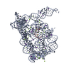
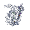
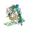
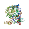
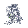
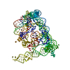
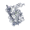
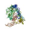
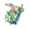
 Movie
Movie Controller


+ Open data

- Basic information
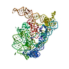
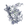
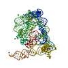
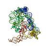
| Entry | Database: PDB / ID: 3eog | ||||||
|---|---|---|---|---|---|---|---|
| Title | Co-crystallization showing exon recognition by a group II intron | ||||||
 Components Components |
| ||||||
 Keywords Keywords | RNA / Ribonucleic acid / Intron / Group II / exon | ||||||
| Function / homology | : / RNA / RNA (> 10) / RNA (> 100) Function and homology information Function and homology information | ||||||
| Method |  X-RAY DIFFRACTION / SYNCHROTRON / MOLECULAR REPLACEMENT / Resolution: 3.391 Å X-RAY DIFFRACTION / SYNCHROTRON / MOLECULAR REPLACEMENT / Resolution: 3.391 Å | ||||||
 Authors Authors | Toor, N. / Rajashankar, K. / Keating, K.S. / Pyle, A.M. | ||||||
 Citation Citation | Journal: Nat.Struct.Mol.Biol. / Year: 2008 Title: Structural basis for exon recognition by a group II intron. Authors: Toor, N. / Rajashankar, K. / Keating, K.S. / Pyle, A.M. | ||||||
| History |
|
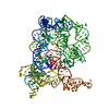
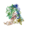
- Structure visualization
| Structure viewer | Molecule: MolmilJmol/JSmol |
|---|
- Downloads & links
-Download
| PDBx/mmCIF format | 3eog.cif.gz | 436.3 KB | Display | PDBx/mmCIF format |
|---|---|---|---|---|
| PDB format | pdb3eog.ent.gz | 355.3 KB | Display | PDB format |
| PDBx/mmJSON format | 3eog.json.gz | Tree view | PDBx/mmJSON format | |
| Others |  Other downloads Other downloads |
-Validation report
| Arichive directory | https://data.pdbj.org/pub/pdb/validation_reports/eo/3eogftp://data.pdbj.org/pub/pdb/validation_reports/eo/3eog | HTTPS FTP |
|---|
-Related structure data


-Links
 PDBj
PDBj


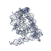
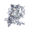
- Assembly
| Deposited unit | 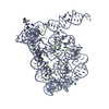
| ||||||||
|---|---|---|---|---|---|---|---|---|---|
| 1 |
| ||||||||
| Unit cell |
|
-Components
| #1: RNA chain | Mass: 133652.156 Da / Num. of mol.: 1 / Source method: obtained synthetically Details: Oceanobacillus iheyensis sequence modified using PCR mutagenesis | ||||
|---|---|---|---|---|---|
| #2: RNA chain | Mass: 1838.117 Da / Num. of mol.: 1 / Source method: obtained synthetically / Details: Ligated exon construct | ||||
| #3: Chemical | ChemComp-MG /   Mass: 24.305 Da / Num. of mol.: 31 / Source method: obtained synthetically / Formula: Mg Mass: 24.305 Da / Num. of mol.: 31 / Source method: obtained synthetically / Formula: Mg#4: Chemical | ChemComp-K /   Mass: 39.098 Da / Num. of mol.: 11 / Source method: obtained synthetically / Formula: K Mass: 39.098 Da / Num. of mol.: 11 / Source method: obtained synthetically / Formula: K#5: Water | ChemComp-HOH / |  Mass: 18.015 Da / Num. of mol.: 15 / Source method: isolated from a natural source / Formula: H2O Mass: 18.015 Da / Num. of mol.: 15 / Source method: isolated from a natural source / Formula: H2O |
-Experimental details
-Experiment
| Experiment | Method: X-RAY DIFFRACTION / Number of used crystals: 1 |
|---|
- Sample preparation
| Crystal | Density Matthews: 3.53 Å3/Da / Density % sol: 65.15 % | ||||||||||||||||||||||||||||||||||||
|---|---|---|---|---|---|---|---|---|---|---|---|---|---|---|---|---|---|---|---|---|---|---|---|---|---|---|---|---|---|---|---|---|---|---|---|---|---|
| Crystal grow | Temperature: 303 K / pH: 7 Details: 6% PEG 3350, 150 mM magnesium acetate, 350 mM KCL, 50 mM Na-HEPES pH 7.0, 0.5 mM spermine, VAPOR DIFFUSION, SITTING DROP, temperature 303K | ||||||||||||||||||||||||||||||||||||
| Components of the solutions |
|
-Data collection
| Diffraction | Mean temperature: 100 K |
|---|---|
| Diffraction source | Source: SYNCHROTRON / Site: APS  / Beamline: 24-ID-C / Wavelength: 0.9795 / Beamline: 24-ID-C / Wavelength: 0.9795 |
| Detector | Type: ADSC QUANTUM 315 / Detector: CCD / Date: Apr 18, 2008 |
| Radiation | Monochromator: SILICON / Protocol: SINGLE WAVELENGTH / Monochromatic (M) / Laue (L): M / Scattering type: x-ray |
| Radiation wavelength | Wavelength: 0.9795 Å / Relative weight: 1 |
| Reflection | Resolution: 3.391→48.6 Å / Num. obs: 27468 |
- Processing
| Software | Name: PHENIX / Version: (phenix.refine) / Classification: refinement | ||||||||||||||||||||||||||||||||||||||||||||||||||||||||||||||||||||||
|---|---|---|---|---|---|---|---|---|---|---|---|---|---|---|---|---|---|---|---|---|---|---|---|---|---|---|---|---|---|---|---|---|---|---|---|---|---|---|---|---|---|---|---|---|---|---|---|---|---|---|---|---|---|---|---|---|---|---|---|---|---|---|---|---|---|---|---|---|---|---|---|
| Refinement | Method to determine structure: MOLECULAR REPLACEMENT / Resolution: 3.391→48.419 Å / SU ML: 0.52 / σ(F): 0.21 / Phase error: 24.59 / Stereochemistry target values: ML
| ||||||||||||||||||||||||||||||||||||||||||||||||||||||||||||||||||||||
| Solvent computation | Shrinkage radii: 0.9 Å / VDW probe radii: 1.11 Å / Solvent model: FLAT BULK SOLVENT MODEL / Bsol: 37.958 Å2 / ksol: 0.173 e/Å3 | ||||||||||||||||||||||||||||||||||||||||||||||||||||||||||||||||||||||
| Refinement step | Cycle: LAST / Resolution: 3.391→48.419 Å
| ||||||||||||||||||||||||||||||||||||||||||||||||||||||||||||||||||||||
| Refine LS restraints |
| ||||||||||||||||||||||||||||||||||||||||||||||||||||||||||||||||||||||
| LS refinement shell |
| ||||||||||||||||||||||||||||||||||||||||||||||||||||||||||||||||||||||
| Refinement TLS params. | Method: refined / Origin x: 19.2473 Å / Origin y: 86.149 Å / Origin z: 363.1506 Å
| ||||||||||||||||||||||||||||||||||||||||||||||||||||||||||||||||||||||
| Refinement TLS group | Selection details: all |
