 Movie
Movie Controller

[English] 日本語
 Yorodumi
- PDB-7o0h: Structure of the foamy viral protease-reverse transcriptase dRH i... -
+ Open data

- Basic information
| Entry | Database: PDB / ID: 7o0h | ||||||
|---|---|---|---|---|---|---|---|
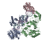
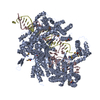
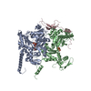
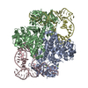
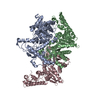
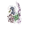
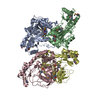
| Title | Structure of the foamy viral protease-reverse transcriptase dRH in complex with ds DNA. | ||||||
 Components Components |
| ||||||
 Keywords Keywords | VIRAL PROTEIN / reverse trancscriptase / complex with dsDNA | ||||||
| Function / homology |  Function and homology information Function and homology informationDNA integration / viral genome integration into host DNA / establishment of integrated proviral latency / virion component / viral penetration into host nucleus / RNA-directed DNA polymerase activity / RNA-DNA hybrid ribonuclease activity / host cell / DNA recombination / aspartic-type endopeptidase activity ...DNA integration / viral genome integration into host DNA / establishment of integrated proviral latency / virion component / viral penetration into host nucleus / RNA-directed DNA polymerase activity / RNA-DNA hybrid ribonuclease activity / host cell / DNA recombination / aspartic-type endopeptidase activity / host cell cytoplasm / DNA-directed DNA polymerase activity / symbiont entry into host cell / host cell nucleus / proteolysis / RNA binding / metal ion binding Similarity search - Function | ||||||
| Biological species |  White-tufted-ear marmoset simian foamy virus White-tufted-ear marmoset simian foamy virussynthetic construct (others) | ||||||
| Method |  X-RAY DIFFRACTION / SYNCHROTRON / MOLECULAR REPLACEMENT / Resolution: 3.09 Å X-RAY DIFFRACTION / SYNCHROTRON / MOLECULAR REPLACEMENT / Resolution: 3.09 Å | ||||||
 Authors Authors | Nowak, E. / Nowacka, M. / Nowotny, M. | ||||||
| Funding support |  Poland, 1items Poland, 1items
| ||||||
 Citation Citation | Journal: J Virol / Year: 2021 Title: Structures of Substrate Complexes of Foamy Viral Protease-Reverse Transcriptase. Authors: Marzena Nowacka / Elżbieta Nowak / Mariusz Czarnocki-Cieciura / Justyna Jackiewicz / Krzysztof Skowronek / Roman H Szczepanowski / Birgitta M Wöhrl / Marcin Nowotny /  Abstract: Reverse transcriptases (RTs) use their DNA polymerase and RNase H activities to catalyze the conversion of single-stranded RNA to double-stranded DNA (dsDNA), a crucial process for the replication of ...Reverse transcriptases (RTs) use their DNA polymerase and RNase H activities to catalyze the conversion of single-stranded RNA to double-stranded DNA (dsDNA), a crucial process for the replication of retroviruses. Foamy viruses (FVs) possess a unique RT, which is a fusion with the protease (PR) domain. The mechanism of substrate binding by this enzyme has been unknown. Here, we report a crystal structure of monomeric full-length marmoset FV (MFV) PR-RT in complex with an RNA/DNA hybrid substrate. We also describe a structure of MFV PR-RT with an RNase H deletion in complex with a dsDNA substrate in which the enzyme forms an asymmetric homodimer. Cryo-electron microscopy reconstruction of the full-length MFV PR-RT-dsDNA complex confirmed the dimeric architecture. These findings represent the first structural description of nucleic acid binding by a foamy viral RT and demonstrate its ability to change its oligomeric state depending on the type of bound nucleic acid. Reverse transcriptases (RTs) are intriguing enzymes converting single-stranded RNA to dsDNA. Their activity is essential for retroviruses, which are divided into two subfamilies differing significantly in their life cycles: and . The latter family is much more ancient and comprises five genera. A unique feature of foamy viral RTs is that they contain N-terminal protease (PR) domains, which are not present in orthoretroviral enzymes. So far, no structural information for full-length foamy viral PR-RT interacting with nucleic substrates has been reported. Here, we present crystal and cryo-electron microscopy structures of marmoset foamy virus (MFV) PR-RT. These structures revealed the mode of binding of RNA/DNA and dsDNA substrates. Moreover, unexpectedly, the structures and biochemical data showed that foamy viral PR-RT can adopt both a monomeric configuration, which is observed in our structures in the presence of an RNA/DNA hybrid, and an asymmetric dimer arrangement, which we observed in the presence of dsDNA. | ||||||
| History |
|
- Structure visualization
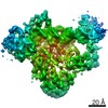
| Structure viewer | Molecule: MolmilJmol/JSmol |
|---|
- Downloads & links
-Download
| PDBx/mmCIF format | 7o0h.cif.gz | 472 KB | Display | PDBx/mmCIF format |
|---|---|---|---|---|
| PDB format | pdb7o0h.ent.gz | 386.6 KB | Display | PDB format |
| PDBx/mmJSON format | 7o0h.json.gz | Tree view | PDBx/mmJSON format | |
| Others |  Other downloads Other downloads |
-Validation report
| Arichive directory | https://data.pdbj.org/pub/pdb/validation_reports/o0/7o0hftp://data.pdbj.org/pub/pdb/validation_reports/o0/7o0h | HTTPS FTP |
|---|
-Related structure data

-Links
 PDBj
PDBj


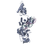
- Assembly
| Deposited unit | 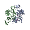
| ||||||||||
|---|---|---|---|---|---|---|---|---|---|---|---|
| 1 |
| ||||||||||
| Unit cell |
|
-Components
| #1: Protein | Mass: 67342.812 Da / Num. of mol.: 2 Source method: isolated from a genetically manipulated source Details: foamy virus Source: (gene. exp.) White-tufted-ear marmoset simian foamy virusGene: pol Production host:  References: UniProt: D5JWV1, RNA-directed DNA polymerase, DNA-directed DNA polymerase, ribonuclease H #2: DNA chain | | Mass: 4612.026 Da / Num. of mol.: 1 / Source method: obtained synthetically / Source: (synth.) synthetic construct (others) #3: DNA chain | | Mass: 3958.571 Da / Num. of mol.: 1 / Source method: obtained synthetically / Source: (synth.) synthetic construct (others) |
|---|
-Experimental details
-Experiment
| Experiment | Method: X-RAY DIFFRACTION / Number of used crystals: 1 |
|---|
- Sample preparation
| Crystal | Density Matthews: 3.47 Å3/Da / Density % sol: 64.58 % |
|---|---|
| Crystal grow | Temperature: 291 K / Method: vapor diffusion, hanging drop / pH: 7 / Details: PEG 3350, DL malic acid |
-Data collection
| Diffraction | Mean temperature: 100 K / Serial crystal experiment: N |
|---|---|
| Diffraction source | Source: SYNCHROTRON / Site: PETRA III, DESY / Beamline: P11 / Wavelength: 1.0332 Å |
| Detector | Type: DECTRIS EIGER2 X 16M / Detector: PIXEL / Date: Dec 14, 2020 |
| Radiation | Protocol: SINGLE WAVELENGTH / Monochromatic (M) / Laue (L): M / Scattering type: x-ray |
| Radiation wavelength | Wavelength: 1.0332 Å / Relative weight: 1 |
| Reflection | Resolution: 3.09→50 Å / Num. obs: 35709 / % possible obs: 99.6 % / Redundancy: 3.8 % / Biso Wilson estimate: 86.18 Å2 / CC1/2: 0.994 / Net I/σ(I): 9.3 |
| Reflection shell | Resolution: 3.09→3.28 Å / Num. unique obs: 5612 / CC1/2: 0.564 |
- Processing
| Software |
| |||||||||||||||||||||||||||||||||||||||||||||||||||||||||||||||||||||||||||||||||||||||||||||||||||||||||||||||||||||||||||||||||||||||||||||||||||||||||||||||||||||||||||||||||||||||||||||||||||||||||||||||||||||||||||||||||
|---|---|---|---|---|---|---|---|---|---|---|---|---|---|---|---|---|---|---|---|---|---|---|---|---|---|---|---|---|---|---|---|---|---|---|---|---|---|---|---|---|---|---|---|---|---|---|---|---|---|---|---|---|---|---|---|---|---|---|---|---|---|---|---|---|---|---|---|---|---|---|---|---|---|---|---|---|---|---|---|---|---|---|---|---|---|---|---|---|---|---|---|---|---|---|---|---|---|---|---|---|---|---|---|---|---|---|---|---|---|---|---|---|---|---|---|---|---|---|---|---|---|---|---|---|---|---|---|---|---|---|---|---|---|---|---|---|---|---|---|---|---|---|---|---|---|---|---|---|---|---|---|---|---|---|---|---|---|---|---|---|---|---|---|---|---|---|---|---|---|---|---|---|---|---|---|---|---|---|---|---|---|---|---|---|---|---|---|---|---|---|---|---|---|---|---|---|---|---|---|---|---|---|---|---|---|---|---|---|---|---|---|---|---|---|---|---|---|---|---|---|---|---|---|---|---|---|
| Refinement | Method to determine structure: MOLECULAR REPLACEMENT Starting model: MFV RT Resolution: 3.09→48.84 Å / SU ML: 0.47 / Cross valid method: THROUGHOUT / σ(F): 1.34 / Phase error: 32.47 / Stereochemistry target values: ML
| |||||||||||||||||||||||||||||||||||||||||||||||||||||||||||||||||||||||||||||||||||||||||||||||||||||||||||||||||||||||||||||||||||||||||||||||||||||||||||||||||||||||||||||||||||||||||||||||||||||||||||||||||||||||||||||||||
| Solvent computation | Shrinkage radii: 0.9 Å / VDW probe radii: 1.11 Å / Solvent model: FLAT BULK SOLVENT MODEL | |||||||||||||||||||||||||||||||||||||||||||||||||||||||||||||||||||||||||||||||||||||||||||||||||||||||||||||||||||||||||||||||||||||||||||||||||||||||||||||||||||||||||||||||||||||||||||||||||||||||||||||||||||||||||||||||||
| Displacement parameters | Biso max: 239.79 Å2 / Biso mean: 104.9667 Å2 / Biso min: 36.74 Å2 | |||||||||||||||||||||||||||||||||||||||||||||||||||||||||||||||||||||||||||||||||||||||||||||||||||||||||||||||||||||||||||||||||||||||||||||||||||||||||||||||||||||||||||||||||||||||||||||||||||||||||||||||||||||||||||||||||
| Refinement step | Cycle: final / Resolution: 3.09→48.84 Å
| |||||||||||||||||||||||||||||||||||||||||||||||||||||||||||||||||||||||||||||||||||||||||||||||||||||||||||||||||||||||||||||||||||||||||||||||||||||||||||||||||||||||||||||||||||||||||||||||||||||||||||||||||||||||||||||||||
| LS refinement shell | Refine-ID: X-RAY DIFFRACTION / Rfactor Rfree error: 0 / Total num. of bins used: 13
| |||||||||||||||||||||||||||||||||||||||||||||||||||||||||||||||||||||||||||||||||||||||||||||||||||||||||||||||||||||||||||||||||||||||||||||||||||||||||||||||||||||||||||||||||||||||||||||||||||||||||||||||||||||||||||||||||
| Refinement TLS params. | Method: refined / Refine-ID: X-RAY DIFFRACTION
| |||||||||||||||||||||||||||||||||||||||||||||||||||||||||||||||||||||||||||||||||||||||||||||||||||||||||||||||||||||||||||||||||||||||||||||||||||||||||||||||||||||||||||||||||||||||||||||||||||||||||||||||||||||||||||||||||
| Refinement TLS group |
|
