 Movie
Movie Controller


+ Open data

- Basic information
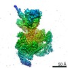
| Entry | Database: EMDB / ID: EMD-12515 | |||||||||
|---|---|---|---|---|---|---|---|---|---|---|
| Title | Vibrio cholerae ParA2-ATPyS-DNA filament | |||||||||
 Map data Map data | ||||||||||
 Sample Sample |
| |||||||||
 Keywords Keywords | ATPase / Chromosome segregation / Bacterial cell division / filament / DNA BINDING PROTEIN | |||||||||
| Function / homology | ParA helix turn helix domain / ParA helix turn helix domain / : / AAA domain / AAA domain / P-loop containing nucleoside triphosphate hydrolase / AAA family ATPase Function and homology information Function and homology information | |||||||||
| Biological species |   Vibrio cholerae (bacteria) / Vibrio cholerae (bacteria) /  Neoarius leptaspis (salmon catfish) Neoarius leptaspis (salmon catfish) | |||||||||
| Method | helical reconstruction / cryo EM / Resolution: 4.5 Å | |||||||||
 Authors Authors | Parker AV / Bergeron JRC | |||||||||
| Funding support |  United Kingdom, 1 items United Kingdom, 1 items
| |||||||||
 Citation Citation | Journal: Nat Commun / Year: 2021 Title: The structure of the bacterial DNA segregation ATPase filament reveals the conformational plasticity of ParA upon DNA binding. Authors: Alexandra V Parker / Daniel Mann / Svetomir B Tzokov / Ling C Hwang / Julien R C Bergeron /  Abstract: The efficient segregation of replicated genetic material is an essential step for cell division. Bacterial cells use several evolutionarily-distinct genome segregation systems, the most common of ...The efficient segregation of replicated genetic material is an essential step for cell division. Bacterial cells use several evolutionarily-distinct genome segregation systems, the most common of which is the type I Par system. It consists of an adapter protein, ParB, that binds to the DNA cargo via interaction with the parS DNA sequence; and an ATPase, ParA, that binds nonspecific DNA and mediates cargo transport. However, the molecular details of how this system functions are not well understood. Here, we report the cryo-EM structure of the Vibrio cholerae ParA2 filament bound to DNA, as well as the crystal structures of this protein in various nucleotide states. These structures show that ParA forms a left-handed filament on DNA, stabilized by nucleotide binding, and that ParA undergoes profound structural rearrangements upon DNA binding and filament assembly. Collectively, our data suggest the structural basis for ParA's cooperative binding to DNA and the formation of high ParA density regions on the nucleoid. | |||||||||
| History |
|
- Structure visualization
| Movie |
Movie viewer |
|---|---|
| Structure viewer | EM map: SurfViewMolmilJmol/JSmol |
| Supplemental images |

- Downloads & links
-EMDB archive
| Map data | emd_12515.map.gz | 13.6 MB | EMDB map data format | |
|---|---|---|---|---|
| Header (meta data) | emd-12515-v30.xmlemd-12515.xml | 18.4 KB 18.4 KB | Display Display | EMDB header |
| Images | 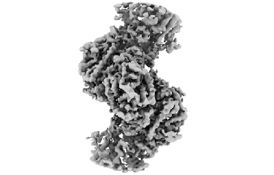 emd_12515.png emd_12515.png | 51.7 KB | ||
| Filedesc metadata | emd-12515.cif.gz | 6.7 KB | ||
| Archive directory |  http://ftp.pdbj.org/pub/emdb/structures/EMD-12515ftp://ftp.pdbj.org/pub/emdb/structures/EMD-12515 http://ftp.pdbj.org/pub/emdb/structures/EMD-12515ftp://ftp.pdbj.org/pub/emdb/structures/EMD-12515 | HTTPS FTP |
-Related structure data
| Related structure data |  7npfMC  7npdC M: atomic model generated by this map C: citing same article ( |
|---|---|
| Similar structure data |


-Links
| EMDB pages | EMDB (EBI/PDBe) / EMDataResource |
|---|
-Map
| File | Download / File: emd_12515.map.gz / Format: CCP4 / Size: 14.6 MB / Type: IMAGE STORED AS FLOATING POINT NUMBER (4 BYTES) | ||||||||||||||||||||||||||||||||||||||||||||||||||||||||||||||||||||
|---|---|---|---|---|---|---|---|---|---|---|---|---|---|---|---|---|---|---|---|---|---|---|---|---|---|---|---|---|---|---|---|---|---|---|---|---|---|---|---|---|---|---|---|---|---|---|---|---|---|---|---|---|---|---|---|---|---|---|---|---|---|---|---|---|---|---|---|---|---|
| Projections & slices | Image control
Images are generated by Spider. generated in cubic-lattice coordinate | ||||||||||||||||||||||||||||||||||||||||||||||||||||||||||||||||||||
| Voxel size | X=Y=Z: 1.04 Å | ||||||||||||||||||||||||||||||||||||||||||||||||||||||||||||||||||||
| Density |
| ||||||||||||||||||||||||||||||||||||||||||||||||||||||||||||||||||||
| Symmetry | Space group: 1 | ||||||||||||||||||||||||||||||||||||||||||||||||||||||||||||||||||||
| Details | EMDB XML:
CCP4 map header:
| ||||||||||||||||||||||||||||||||||||||||||||||||||||||||||||||||||||
 X (Sec.)
 Y (Row.)
 Z (Col.)


-Supplemental data
- Sample components
-Entire : ParA2-ATPgS-DNA
| Entire | Name: ParA2-ATPgS-DNA |
|---|---|
| Components |
|
-Supramolecule #1: ParA2-ATPgS-DNA
| Supramolecule | Name: ParA2-ATPgS-DNA / type: complex / ID: 1 / Parent: 0 / Macromolecule list: #1-#3 |
|---|
-Macromolecule #1: AAA family ATPase
| Macromolecule | Name: AAA family ATPase / type: protein_or_peptide / ID: 1 / Number of copies: 8 / Enantiomer: LEVO |
|---|---|
| Source (natural) | Organism: Vibrio cholerae (bacteria) |
| Molecular weight | Theoretical: 46.440969 KDa |
| Recombinant expression | Organism: |
| Sequence | String: MAMKREQTIE NLYQLAQLTQ QVQADRIEIV LEERRDEHFP PMSKALMETR SGLTRRKLDE AIAKMEEAGH QFTKNNANHY SISLSEAHM LMDAAGVPKF HERKKNNENK PWIINVQNQK GGTGKSMTAV HLAACLALNL DKRYRICLID LDPQGSLRLF L NPQISLAE ...String: MAMKREQTIE NLYQLAQLTQ QVQADRIEIV LEERRDEHFP PMSKALMETR SGLTRRKLDE AIAKMEEAGH QFTKNNANHY SISLSEAHM LMDAAGVPKF HERKKNNENK PWIINVQNQK GGTGKSMTAV HLAACLALNL DKRYRICLID LDPQGSLRLF L NPQISLAE HTNIYSAVDI MLDNVPDGVQ VDTEFLRKNV MLPTQYPNLK TISAFPEDAM FNAEAWQYLS QNQSLDIVRL LK EKLIDKI ASDFDIIMID TGPHVDPLVW NAMYASNALL IPCAAKRLDW ASTVNFFQHL PTVYEMFPED WKGLEFVRLM PTM FEDDNK KQVSVLTEMN YLLGDQVMMA TIPRSRAFET CADTYSTVFD LTVNDFEGGK KTLATAQDAV QKSALELERV LHSH WSSLN QG UniProtKB: AAA family ATPase |
-Macromolecule #2: DNA (49-MER)
| Macromolecule | Name: DNA (49-MER) / type: dna / ID: 2 / Number of copies: 1 / Classification: DNA |
|---|---|
| Source (natural) | Organism: Neoarius leptaspis (salmon catfish) |
| Molecular weight | Theoretical: 15.30217 KDa |
| Sequence | String: (DA)(DA)(DA)(DA)(DA)(DA)(DA)(DA)(DA)(DA) (DA)(DA)(DA)(DA)(DA)(DA)(DA)(DA)(DA)(DA) (DA)(DA)(DA)(DA)(DA)(DA)(DA)(DA)(DA) (DA)(DA)(DA)(DA)(DA)(DA)(DA)(DA)(DA)(DA) (DA) (DA)(DA)(DA)(DA)(DA)(DA)(DA)(DA) (DA) |
-Macromolecule #3: DNA (49-MER)
| Macromolecule | Name: DNA (49-MER) / type: dna / ID: 3 / Number of copies: 1 / Classification: DNA |
|---|---|
| Source (natural) | Organism: Neoarius leptaspis (salmon catfish) |
| Molecular weight | Theoretical: 14.86049 KDa |
| Sequence | String: (DT)(DT)(DT)(DT)(DT)(DT)(DT)(DT)(DT)(DT) (DT)(DT)(DT)(DT)(DT)(DT)(DT)(DT)(DT)(DT) (DT)(DT)(DT)(DT)(DT)(DT)(DT)(DT)(DT) (DT)(DT)(DT)(DT)(DT)(DT)(DT)(DT)(DT)(DT) (DT) (DT)(DT)(DT)(DT)(DT)(DT)(DT)(DT) (DT) |
-Macromolecule #4: MAGNESIUM ION
| Macromolecule | Name: MAGNESIUM ION / type: ligand / ID: 4 / Number of copies: 8 / Formula: MG |
|---|---|
| Molecular weight | Theoretical: 24.305 Da |
-Macromolecule #5: PHOSPHOTHIOPHOSPHORIC ACID-ADENYLATE ESTER
| Macromolecule | Name: PHOSPHOTHIOPHOSPHORIC ACID-ADENYLATE ESTER / type: ligand / ID: 5 / Number of copies: 8 / Formula: AGS |
|---|---|
| Molecular weight | Theoretical: 523.247 Da |
| Chemical component information |  ChemComp-AGS: |
-Experimental details
-Structure determination
| Method | cryo EM |
|---|---|
 Processing Processing | helical reconstruction |
| Aggregation state | filament |
-Sample preparation
| Buffer | pH: 7 |
|---|---|
| Vitrification | Cryogen name: ETHANE |
- Electron microscopy
| Microscope | FEI TITAN KRIOS |
|---|---|
| Image recording | Film or detector model: GATAN K2 SUMMIT (4k x 4k) / Average electron dose: 52.02 e/Å2 |
| Electron beam | Acceleration voltage: 300 kV / Electron source:  FIELD EMISSION GUN FIELD EMISSION GUN |
| Electron optics | Illumination mode: FLOOD BEAM / Imaging mode: BRIGHT FIELD |
| Experimental equipment |  Model: Titan Krios / Image courtesy: FEI Company |
-Image processing
| Final reconstruction | Applied symmetry - Helical parameters - Δz: 28.68 Å Applied symmetry - Helical parameters - Δ&Phi: -80.57 ° Applied symmetry - Helical parameters - Axial symmetry: C1 (asymmetric) Resolution.type: BY AUTHOR / Resolution: 4.5 Å / Resolution method: FSC 0.143 CUT-OFF / Number images used: 182997 |
|---|---|
| CTF correction | Type: PHASE FLIPPING AND AMPLITUDE CORRECTION |
| Startup model | Type of model: PDB ENTRY PDB model - PDB ID: |
| Final angle assignment | Type: NOT APPLICABLE |

-Atomic model buiding 1
| Refinement | Protocol: OTHER |
|---|---|
| Output model | PDB-7npf: |
