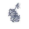
Mass: 132344.812 Da / Num. of mol.: 4 / Mutation: E178Q Source method: isolated from a genetically manipulated source Source: (gene. exp.) Kluyveromyces lactis (yeast) / Gene: KLLA0F22385g / Production host: Escherichia coli (E. coli) / References: UniProt: Q6CJ09
Sequence details
E178Q IS AN ACTIVE SITE MUTATION. AMINO ACID SEQUENCE CORRESPONDING TO RESIDUES 1-1245 IN UNIPROT ...E178Q IS AN ACTIVE SITE MUTATION. AMINO ACID SEQUENCE CORRESPONDING TO RESIDUES 1-1245 IN UNIPROT DATABASE ENTRY Q6CJ09, PLUS A C-TERMINAL EXPRESSION TAG (RESIDUES 1246-1253), WAS USED FOR CRYSTALLIZATION. MOST OF THE RESIDUES FROM RESIDUE 463 TO 503, AND FROM RESIDUE 984 TO 1082 WERE MISSING IN THE COORDINATES DUE TO LACK OF ELECTRON DENSITY. SEVERAL RESIDUES WITHIN THESE RANGES WERE MODELED AS POLY-ALA SEGMENTS, AND ARE REPRESENTED AS UNK RESIDUES 469-487 AND 1038-1052 OF CHAIN A, UNK RESIDUES 469-487 AND 1036-1052 OF CHAIN B, UNK RESIDUES 469-487 AND 1038-1054 OF CHAIN C, AND UNK RESIDUES 469-487 OF CHAIN D. THESE RESIDUE NUMBERS ARE ARBITRARILY ASSIGNED. IT IS EXPECTED THAT RESIDUES 469-487 BELONG TO THE 463-503 SEGMENT, AND RESIDUES 1036-1054 BELONG TO THE 984-1082 SEGMENT.
-
Experimental details
-
Experiment
Experiment
Method: X-RAY DIFFRACTION / Number of used crystals: 1
-
Sample preparation
Crystal
Density Matthews: 3.5 Å3/Da / Density % sol: 64.88 %
Crystal grow
Temperature: 277 K / Method: vapor diffusion, sitting drop / pH: 8.5 Details: 0.1M Tris, 0.9M LiCl, 13.5% PEG 6000, pH 8.5, VAPOR DIFFUSION, SITTING DROP, temperature 277K
Method to determine structure: MOLECULAR REPLACEMENT / Resolution: 2.9→30 Å / Cor.coef. Fo:Fc: 0.909 / Cor.coef. Fo:Fc free: 0.857 / SU B: 41.844 / SU ML: 0.36 / Cross valid method: THROUGHOUT / ESU R Free: 0.437 / Stereochemistry target values: MAXIMUM LIKELIHOOD / Details: HYDROGENS HAVE BEEN ADDED IN THE RIDING POSITIONS
Rfactor
Num. reflection
% reflection
Selection details
Rfree
0.30476
7790
5 %
RANDOM
Rwork
0.24125
-
-
-
obs
0.24441
146829
97.22 %
-
all
-
151028
-
-
Solvent computation
Ion probe radii: 0.8 Å / Shrinkage radii: 0.8 Å / VDW probe radii: 1.4 Å / Solvent model: MASK
Displacement parameters
Biso mean: 71.675 Å2
Baniso -1
Baniso -2
Baniso -3
1-
2.37 Å2
3.39 Å2
3.59 Å2
2-
-
0.81 Å2
2.86 Å2
3-
-
-
2.33 Å2
Refinement step
Cycle: LAST / Resolution: 2.9→30 Å
Protein
Nucleic acid
Ligand
Solvent
Total
Num. atoms
34009
0
0
0
34009
Refine LS restraints
Refine-ID
Type
Dev ideal
Dev ideal target
Number
X-RAY DIFFRACTION
r_bond_refined_d
0.016
0.022
34786
X-RAY DIFFRACTION
r_bond_other_d
X-RAY DIFFRACTION
r_angle_refined_deg
1.679
1.963
46933
X-RAY DIFFRACTION
r_angle_other_deg
X-RAY DIFFRACTION
r_dihedral_angle_1_deg
7.086
5
4151
X-RAY DIFFRACTION
r_dihedral_angle_2_deg
38.752
24.118
1673
X-RAY DIFFRACTION
r_dihedral_angle_3_deg
21.746
15
6238
X-RAY DIFFRACTION
r_dihedral_angle_4_deg
18.747
15
199
X-RAY DIFFRACTION
r_chiral_restr
0.109
0.2
5121
X-RAY DIFFRACTION
r_gen_planes_refined
0.007
0.021
26170
X-RAY DIFFRACTION
r_gen_planes_other
X-RAY DIFFRACTION
r_nbd_refined
X-RAY DIFFRACTION
r_nbd_other
X-RAY DIFFRACTION
r_nbtor_refined
X-RAY DIFFRACTION
r_nbtor_other
X-RAY DIFFRACTION
r_xyhbond_nbd_refined
X-RAY DIFFRACTION
r_xyhbond_nbd_other
X-RAY DIFFRACTION
r_metal_ion_refined
X-RAY DIFFRACTION
r_metal_ion_other
X-RAY DIFFRACTION
r_symmetry_vdw_refined
X-RAY DIFFRACTION
r_symmetry_vdw_other
X-RAY DIFFRACTION
r_symmetry_hbond_refined
X-RAY DIFFRACTION
r_symmetry_hbond_other
X-RAY DIFFRACTION
r_symmetry_metal_ion_refined
X-RAY DIFFRACTION
r_symmetry_metal_ion_other
X-RAY DIFFRACTION
r_mcbond_it
0.976
1.5
20930
X-RAY DIFFRACTION
r_mcbond_other
X-RAY DIFFRACTION
r_mcangle_it
1.809
2
33714
X-RAY DIFFRACTION
r_scbond_it
2.412
3
13856
X-RAY DIFFRACTION
r_scangle_it
3.906
4.5
13219
X-RAY DIFFRACTION
r_rigid_bond_restr
1.333
3
34637
X-RAY DIFFRACTION
r_sphericity_free
X-RAY DIFFRACTION
r_sphericity_bonded
LS refinement shell
Resolution: 2.9→3.055 Å / Total num. of bins used: 10
In the structure databanks used in Yorodumi, some data are registered as the other names, "COVID-19 virus" and "2019-nCoV". Here are the details of the virus and the list of structure data.
Jan 31, 2019. EMDB accession codes are about to change! (news from PDBe EMDB page)
EMDB accession codes are about to change! (news from PDBe EMDB page)
The allocation of 4 digits for EMDB accession codes will soon come to an end. Whilst these codes will remain in use, new EMDB accession codes will include an additional digit and will expand incrementally as the available range of codes is exhausted. The current 4-digit format prefixed with “EMD-” (i.e. EMD-XXXX) will advance to a 5-digit format (i.e. EMD-XXXXX), and so on. It is currently estimated that the 4-digit codes will be depleted around Spring 2019, at which point the 5-digit format will come into force.
The EM Navigator/Yorodumi systems omit the EMD- prefix.
Related info.:Q: What is EMD? / ID/Accession-code notation in Yorodumi/EM Navigator
Yorodumi is a browser for structure data from EMDB, PDB, SASBDB, etc.
This page is also the successor to EM Navigator detail page, and also detail information page/front-end page for Omokage search.
The word "yorodu" (or yorozu) is an old Japanese word meaning "ten thousand". "mi" (miru) is to see.
Related info.:EMDB / PDB / SASBDB / Comparison of 3 databanks / Yorodumi Search / Aug 31, 2016. New EM Navigator & Yorodumi / Yorodumi Papers / Jmol/JSmol / Function and homology information / Changes in new EM Navigator and Yorodumi
 Movie
Movie Controller

 Yorodumi
 Open data
 Basic information
 Components
 Keywords
 Function and homology information
 Kluyveromyces lactis (yeast)
 X-RAY DIFFRACTION /
  Authors
 Citation
 Structure visualization
 Downloads & links
 Other downloads


 PDBj
PDBj Assembly
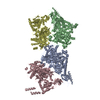


 Sample preparation
 / Beamline: X29A / Wavelength: 1.075 Å
 Processing