+ Open data
- Basic information
| Entry | Database: EMDB / ID: EMD-11665 | ||||||||||||
|---|---|---|---|---|---|---|---|---|---|---|---|---|---|
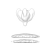
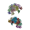
| Title | Structure of GP in EBOV VP40-GP VLPs | ||||||||||||
 Map data Map data | Structure of GP in EBOV VP40-GP VLPs | ||||||||||||
 Sample Sample |
| ||||||||||||
| Biological species |   | ||||||||||||
| Method | subtomogram averaging / cryo EM / Resolution: 12.8 Å | ||||||||||||
 Authors Authors | Wan W / Clarke M / Norris M / Kolesnikova L / Koehler A / Bornholdt ZA / Becker S / Saphire EO / Briggs JAG | ||||||||||||
| Funding support | European Union,  Germany, 3 items Germany, 3 items
| ||||||||||||
 Citation Citation |  Journal: Elife / Year: 2020 Journal: Elife / Year: 2020Title: Ebola and Marburg virus matrix layers are locally ordered assemblies of VP40 dimers. Authors: William Wan / Mairi Clarke / Michael J Norris / Larissa Kolesnikova / Alexander Koehler / Zachary A Bornholdt / Stephan Becker / Erica Ollmann Saphire / John Ag Briggs /    Abstract: Filoviruses such as Ebola and Marburg virus bud from the host membrane as enveloped virions. This process is achieved by the matrix protein VP40. When expressed alone, VP40 induces budding of ...Filoviruses such as Ebola and Marburg virus bud from the host membrane as enveloped virions. This process is achieved by the matrix protein VP40. When expressed alone, VP40 induces budding of filamentous virus-like particles, suggesting that localization to the plasma membrane, oligomerization into a matrix layer, and generation of membrane curvature are intrinsic properties of VP40. There has been no direct information on the structure of VP40 matrix layers within viruses or virus-like particles. We present structures of Ebola and Marburg VP40 matrix layers in intact virus-like particles, and within intact Marburg viruses. VP40 dimers assemble extended chains via C-terminal domain interactions. These chains stack to form 2D matrix lattices below the membrane surface. These lattices form a patchwork assembly across the membrane and suggesting that assembly may begin at multiple points. Our observations define the structure and arrangement of the matrix protein layer that mediates formation of filovirus particles. | ||||||||||||
| History |
|
- Structure visualization
| Movie |
 Movie viewer Movie viewer |
|---|---|
| Structure viewer | EM map:  SurfView SurfView Molmil Molmil Jmol/JSmol Jmol/JSmol |
| Supplemental images |
- Downloads & links
-EMDB archive
| Map data |  emd_11665.map.gz emd_11665.map.gz | 23.8 MB |  EMDB map data format EMDB map data format | |
|---|---|---|---|---|
| Header (meta data) |  emd-11665-v30.xml emd-11665-v30.xml emd-11665.xml emd-11665.xml | 11.3 KB 11.3 KB | Display Display |  EMDB header EMDB header |
| Images |  emd_11665.png emd_11665.png | 18 KB | ||
| Archive directory |  http://ftp.pdbj.org/pub/emdb/structures/EMD-11665 http://ftp.pdbj.org/pub/emdb/structures/EMD-11665 ftp://ftp.pdbj.org/pub/emdb/structures/EMD-11665 ftp://ftp.pdbj.org/pub/emdb/structures/EMD-11665 | HTTPS FTP |
-Validation report
| Summary document |  emd_11665_validation.pdf.gz emd_11665_validation.pdf.gz | 206.8 KB | Display |  EMDB validaton report EMDB validaton report |
|---|---|---|---|---|
| Full document |  emd_11665_full_validation.pdf.gz emd_11665_full_validation.pdf.gz | 205.9 KB | Display | |
| Data in XML |  emd_11665_validation.xml.gz emd_11665_validation.xml.gz | 5.9 KB | Display | |
| Arichive directory |  https://ftp.pdbj.org/pub/emdb/validation_reports/EMD-11665 https://ftp.pdbj.org/pub/emdb/validation_reports/EMD-11665 ftp://ftp.pdbj.org/pub/emdb/validation_reports/EMD-11665 ftp://ftp.pdbj.org/pub/emdb/validation_reports/EMD-11665 | HTTPS FTP |
-Related structure data
- Links
Links
| EMDB pages |  EMDB (EBI/PDBe) / EMDB (EBI/PDBe) /  EMDataResource EMDataResource |
|---|
- Map
Map
| File |  Download / File: emd_11665.map.gz / Format: CCP4 / Size: 27 MB / Type: IMAGE STORED AS FLOATING POINT NUMBER (4 BYTES) Download / File: emd_11665.map.gz / Format: CCP4 / Size: 27 MB / Type: IMAGE STORED AS FLOATING POINT NUMBER (4 BYTES) | ||||||||||||||||||||||||||||||||||||||||||||||||||||||||||||||||||||
|---|---|---|---|---|---|---|---|---|---|---|---|---|---|---|---|---|---|---|---|---|---|---|---|---|---|---|---|---|---|---|---|---|---|---|---|---|---|---|---|---|---|---|---|---|---|---|---|---|---|---|---|---|---|---|---|---|---|---|---|---|---|---|---|---|---|---|---|---|---|
| Annotation | Structure of GP in EBOV VP40-GP VLPs | ||||||||||||||||||||||||||||||||||||||||||||||||||||||||||||||||||||
| Projections & slices | Image control
Images are generated by Spider. | ||||||||||||||||||||||||||||||||||||||||||||||||||||||||||||||||||||
| Voxel size | X=Y=Z: 1.78 Å | ||||||||||||||||||||||||||||||||||||||||||||||||||||||||||||||||||||
| Density |
| ||||||||||||||||||||||||||||||||||||||||||||||||||||||||||||||||||||
| Symmetry | Space group: 1 | ||||||||||||||||||||||||||||||||||||||||||||||||||||||||||||||||||||
| Details | EMDB XML:
CCP4 map header:
| ||||||||||||||||||||||||||||||||||||||||||||||||||||||||||||||||||||
-Supplemental data
- Sample components
-Entire : Ebola virus - Mayinga, Zaire, 1976
| Entire | Name:  |
|---|---|
| Components |
|
-Supramolecule #1: Ebola virus - Mayinga, Zaire, 1976
| Supramolecule | Name: Ebola virus - Mayinga, Zaire, 1976 / type: virus / ID: 1 / Parent: 0 / Macromolecule list: all / NCBI-ID: 128952 / Sci species name: Ebola virus - Mayinga, Zaire, 1976 / Virus type: VIRUS-LIKE PARTICLE / Virus isolate: SPECIES / Virus enveloped: Yes / Virus empty: Yes |
|---|---|
| Host system | Organism:  Homo sapiens (human) Homo sapiens (human) |
-Macromolecule #1: GP
| Macromolecule | Name: GP / type: protein_or_peptide / ID: 1 / Enantiomer: LEVO |
|---|---|
| Source (natural) | Organism:  |
| Recombinant expression | Organism:  Homo sapiens (human) Homo sapiens (human) |
| Sequence | String: MGVTGILQLP RDRFKRTSFF LWVIILFQRT FSIPLGVIHN STLQVSDVDK LVCRDKLSST NQLRSVGLN LEGNGVATDV PSATKRWGFR SGVPPKVVNY EAGEWAENCY NLEIKKPDGS E CLPAAPDG IRGFPRCRYV HKVSGTGPCA GDFAFHKEGA FFLYDRLAST ...String: MGVTGILQLP RDRFKRTSFF LWVIILFQRT FSIPLGVIHN STLQVSDVDK LVCRDKLSST NQLRSVGLN LEGNGVATDV PSATKRWGFR SGVPPKVVNY EAGEWAENCY NLEIKKPDGS E CLPAAPDG IRGFPRCRYV HKVSGTGPCA GDFAFHKEGA FFLYDRLAST VIYRGTTFAE GV VAFLILP QAKKDFFSSH PLREPVNATE DPSSGYYSTT IRYQATGFGT NETEYLFEVD NLT YVQLES RFTPQFLLQL NETIYTSGKR SNTTGKLIWK VNPEIDTTIG EWAFWETKKN LTRK IRSEE LSFTVVSNGA KNISGQSPAR TSSDPGTNTT TEDHKIMASE NSSAMVQVHS QGREA AVSH LTTLATISTS PQSLTTKPGP DNSTHNTPVY KLDISEATQV EQHHRRTDND STASDT PSA TTAAGPPKAE NTNTSKSTDF LDPATTTSPQ NHSETAGNNN THHQDTGEES ASSGKLG LI TNTIAGVAGL ITGGRRTRRE AIVNAQPKCN PNLHYWTTQD EGAAIGLAWI PYFGPAAE G IYIEGLMHNQ DGLICGLRQL ANETTQALQL FLRATTELRT FSILNRKAID FLLQRWGGT CHILGPDCCI EPHDWTKNIT DKIDQIIHDF VDKTLPDQGD NDNWWTGWRQ WIPAGIGVTG VIIAVIALF CICKFVF |
-Experimental details
-Structure determination
| Method | cryo EM |
|---|---|
 Processing Processing | subtomogram averaging |
| Aggregation state | filament |
- Sample preparation
| Buffer | pH: 7.4 |
|---|---|
| Vitrification | Cryogen name: ETHANE |
- Electron microscopy
| Microscope | FEI TITAN KRIOS |
|---|---|
| Image recording | Film or detector model: GATAN K2 QUANTUM (4k x 4k) / Average electron dose: 2.4 e/Å2 |
| Electron beam | Acceleration voltage: 300 kV / Electron source:  FIELD EMISSION GUN FIELD EMISSION GUN |
| Electron optics | Illumination mode: FLOOD BEAM / Imaging mode: BRIGHT FIELD |
| Experimental equipment |  Model: Titan Krios / Image courtesy: FEI Company |
- Image processing
| Final reconstruction | Applied symmetry - Point group: C3 (3 fold cyclic) / Resolution.type: BY AUTHOR / Resolution: 12.8 Å / Resolution method: FSC 0.143 CUT-OFF / Software - Name: AV3 / Number subtomograms used: 11188 | ||||||
|---|---|---|---|---|---|---|---|
| Extraction | Number tomograms: 55 / Number images used: 164205 | ||||||
| CTF correction | Software:
| ||||||
| Final angle assignment | Type: OTHER / Software: (Name: TOM, AV3) / Details: Constrained Cross Correlation |
 Movie
Movie Controller


 Z (Sec.)
 Y (Row.)
 X (Col.)


