 Movie
Movie Controller

[English] 日本語
 Yorodumi
- EMDB-11266: E2 core of the fungal Pyruvate dehydrogenase complex with flexibl... -
+ Open data

- Basic information
| Entry | Database: EMDB / ID: EMD-11266 | ||||||||||||||||||
|---|---|---|---|---|---|---|---|---|---|---|---|---|---|---|---|---|---|---|---|
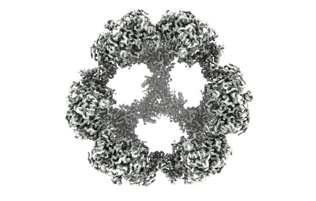
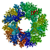
| Title | E2 core of the fungal Pyruvate dehydrogenase complex with flexible/oversym. periphery, structured core and oversym. interior | ||||||||||||||||||
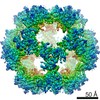
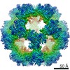
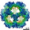
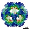
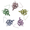
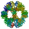
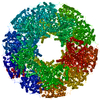
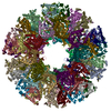
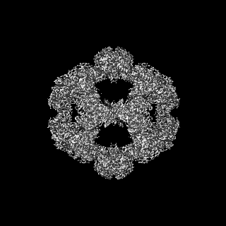
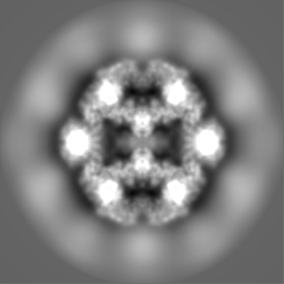
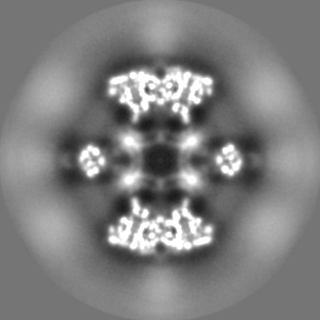
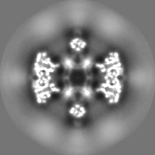
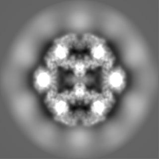
 Map data Map data | Fungal PDC (N. crassa). Endogenous preparation-E1 E2 E3 PX. Enforced symmetry: I2. Periphery is flexible/oversym. Core is structured. Interior is oversym. | ||||||||||||||||||
 Sample Sample |
| ||||||||||||||||||
| Biological species |  Neurospora crassa (fungus) Neurospora crassa (fungus) | ||||||||||||||||||
| Method | single particle reconstruction / cryo EM / Resolution: 4.1 Å | ||||||||||||||||||
 Authors Authors | Forsberg BO / Lindahl E / Aibara S / Howard RJ / Mortezaei N | ||||||||||||||||||
| Funding support |  Sweden, European Union, 5 items Sweden, European Union, 5 items
| ||||||||||||||||||
 Citation Citation | Journal: Nat Commun / Year: 2020 Title: Arrangement and symmetry of the fungal E3BP-containing core of the pyruvate dehydrogenase complex. Authors: B O Forsberg / S Aibara / R J Howard / N Mortezaei / E Lindahl /  Abstract: The pyruvate dehydrogenase complex (PDC) is a multienzyme complex central to aerobic respiration, connecting glycolysis to mitochondrial oxidation of pyruvate. Similar to the E3-binding protein (E3BP) ...The pyruvate dehydrogenase complex (PDC) is a multienzyme complex central to aerobic respiration, connecting glycolysis to mitochondrial oxidation of pyruvate. Similar to the E3-binding protein (E3BP) of mammalian PDC, PX selectively recruits E3 to the fungal PDC, but its divergent sequence suggests a distinct structural mechanism. Here, we report reconstructions of PDC from the filamentous fungus Neurospora crassa by cryo-electron microscopy, where we find protein X (PX) interior to the PDC core as opposed to substituting E2 core subunits as in mammals. Steric occlusion limits PX binding, resulting in predominantly tetrahedral symmetry, explaining previous observations in Saccharomyces cerevisiae. The PX-binding site is conserved in (and specific to) fungi, and complements possible C-terminal binding motifs in PX that are absent in mammalian E3BP. Consideration of multiple symmetries thus reveals a differential structural basis for E3BP-like function in fungal PDC. | ||||||||||||||||||
| History |
|
- Structure visualization
| Movie |
 Movie viewer Movie viewer |
|---|---|
| Structure viewer | EM map: SurfViewMolmilJmol/JSmol |
| Supplemental images |
- Downloads & links
-EMDB archive
| Map data | emd_11266.map.gz | 93.1 MB | EMDB map data format | |
|---|---|---|---|---|
| Header (meta data) | emd-11266-v30.xmlemd-11266.xml | 14.2 KB 14.2 KB | Display Display | EMDB header |
| FSC (resolution estimation) | emd_11266_fsc.xml | 12.9 KB | Display | FSC data file |
| Images |  emd_11266.png emd_11266.png | 213.7 KB | ||
| Others | emd_11266_half_map_1.map.gzemd_11266_half_map_2.map.gz | 96.9 MB 96.9 MB | ||
| Archive directory |  http://ftp.pdbj.org/pub/emdb/structures/EMD-11266ftp://ftp.pdbj.org/pub/emdb/structures/EMD-11266 http://ftp.pdbj.org/pub/emdb/structures/EMD-11266ftp://ftp.pdbj.org/pub/emdb/structures/EMD-11266 | HTTPS FTP |
-Related structure data
| Related structure data |  6zlmC  6zloC C: citing same article ( |
|---|---|
| Similar structure data | |
| EM raw data | EMPIAR-10489 (Title: Native Pyruvate Dehydrogenase Complex from Neurospora crassa Data size: 305.5 Data #1: Aligned and dose-weighted micrographs of native N. crassa Pyruvate dehydrogenase [micrographs - single frame]) |

-Links
| EMDB pages | EMDB (EBI/PDBe) / EMDataResource |
|---|
-Map
| File | Download / File: emd_11266.map.gz / Format: CCP4 / Size: 125 MB / Type: IMAGE STORED AS FLOATING POINT NUMBER (4 BYTES) | ||||||||||||||||||||||||||||||||||||||||||||||||||||||||||||||||||||
|---|---|---|---|---|---|---|---|---|---|---|---|---|---|---|---|---|---|---|---|---|---|---|---|---|---|---|---|---|---|---|---|---|---|---|---|---|---|---|---|---|---|---|---|---|---|---|---|---|---|---|---|---|---|---|---|---|---|---|---|---|---|---|---|---|---|---|---|---|---|
| Annotation | Fungal PDC (N. crassa). Endogenous preparation-E1 E2 E3 PX. Enforced symmetry: I2. Periphery is flexible/oversym. Core is structured. Interior is oversym. | ||||||||||||||||||||||||||||||||||||||||||||||||||||||||||||||||||||
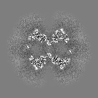
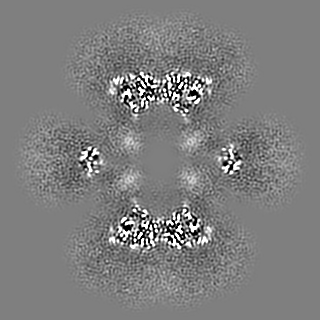
| Projections & slices | Image control
Images are generated by Spider. | ||||||||||||||||||||||||||||||||||||||||||||||||||||||||||||||||||||
| Voxel size | X=Y=Z: 1.25 Å | ||||||||||||||||||||||||||||||||||||||||||||||||||||||||||||||||||||
| Density |
| ||||||||||||||||||||||||||||||||||||||||||||||||||||||||||||||||||||
| Symmetry | Space group: 1 | ||||||||||||||||||||||||||||||||||||||||||||||||||||||||||||||||||||
| Details | EMDB XML:
CCP4 map header:
| ||||||||||||||||||||||||||||||||||||||||||||||||||||||||||||||||||||
 Z (Sec.)
 Y (Row.)
 X (Col.)


-Supplemental data
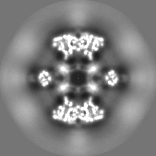
-Half map: Half-map 1. Fungal PDC (N. crassa). Endogenous preparation-E1...
| File | emd_11266_half_map_1.map | ||||||||||||
|---|---|---|---|---|---|---|---|---|---|---|---|---|---|
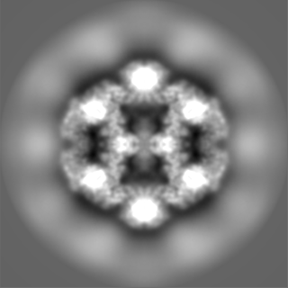
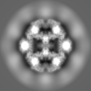
| Annotation | Half-map 1. Fungal PDC (N. crassa). Endogenous preparation-E1 E2 E3 PX. Enforced symmetry: I2. Periphery is flexible/oversym. Core is structured. Interior is oversym. | ||||||||||||
| Projections & Slices |
| ||||||||||||
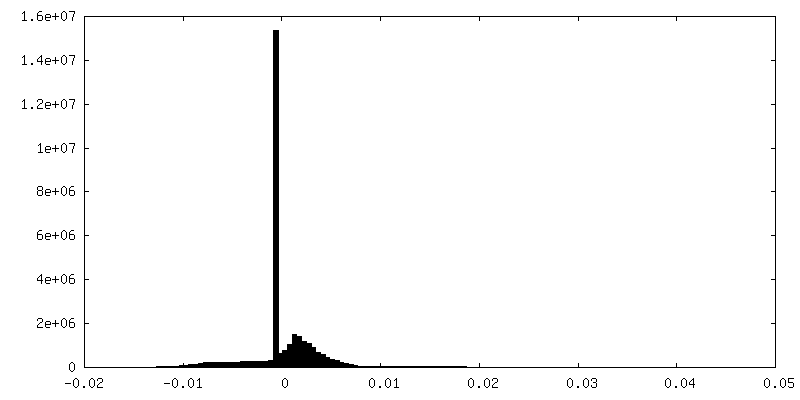
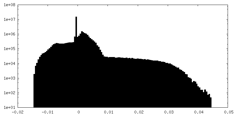
| Density Histograms |

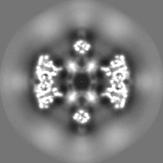
-Half map: Half-map 2. Fungal PDC (N. crassa). Endogenous preparation-E1...
| File | emd_11266_half_map_2.map | ||||||||||||
|---|---|---|---|---|---|---|---|---|---|---|---|---|---|
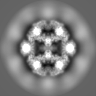
| Annotation | Half-map 2. Fungal PDC (N. crassa). Endogenous preparation-E1 E2 E3 PX. Enforced symmetry: I2. Periphery is flexible/oversym. Core is structured. Interior is oversym. | ||||||||||||
| Projections & Slices |
| ||||||||||||
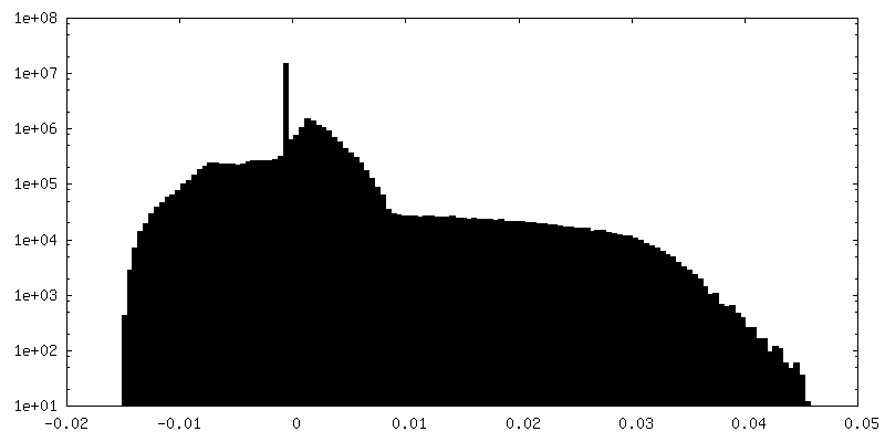
| Density Histograms |


- Sample components
-Entire : endogenous pyruvate dehydrogenase complex form Neurospora crassa
| Entire | Name: endogenous pyruvate dehydrogenase complex form Neurospora crassa |
|---|---|
| Components |
|
-Supramolecule #1: endogenous pyruvate dehydrogenase complex form Neurospora crassa
| Supramolecule | Name: endogenous pyruvate dehydrogenase complex form Neurospora crassa type: complex / ID: 1 / Parent: 0 Details: Catalytic (C-terminal) domain of Dihydrolipoyllysine-residue acetyltransferase (E2-component of pyruvate dehydrogenase complex) |
|---|---|
| Source (natural) | Organism: Neurospora crassa (fungus) / Organelle: mitochondria |
| Molecular weight | Theoretical: 7 MDa |
-Experimental details
-Structure determination
| Method | cryo EM |
|---|---|
 Processing Processing | single particle reconstruction |
| Aggregation state | particle |
-Sample preparation
| Concentration | 3 mg/mL |
|---|---|
| Buffer | pH: 7.5 |
| Grid | Model: Quantifoil R1.2/1.3 / Material: COPPER / Mesh: 300 / Support film - Material: CARBON / Support film - topology: HOLEY / Pretreatment - Type: GLOW DISCHARGE |
| Vitrification | Cryogen name: ETHANE / Chamber humidity: 100 % / Chamber temperature: 277 K / Instrument: FEI VITROBOT MARK IV |
- Electron microscopy
| Microscope | FEI TALOS ARCTICA |
|---|---|
| Image recording | Film or detector model: FEI FALCON II (4k x 4k) / Detector mode: COUNTING / Average electron dose: 35.0 e/Å2 |
| Electron beam | Acceleration voltage: 200 kV / Electron source:  FIELD EMISSION GUN FIELD EMISSION GUN |
| Electron optics | Illumination mode: FLOOD BEAM / Imaging mode: BRIGHT FIELD |
| Experimental equipment |  Model: Talos Arctica / Image courtesy: FEI Company |
-Image processing
| Final reconstruction | Applied symmetry - Point group: I (icosahedral) / Resolution.type: BY AUTHOR / Resolution: 4.1 Å / Resolution method: FSC 0.143 CUT-OFF / Number images used: 69538 |
|---|---|
| Initial angle assignment | Type: MAXIMUM LIKELIHOOD |
| Final angle assignment | Type: MAXIMUM LIKELIHOOD |
| FSC plot (resolution estimation) | 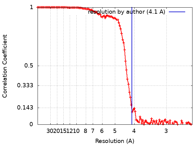 |
