 Movie
Movie Controller

[English] 日本語
 Yorodumi
- EMDB-7610: Atomic Structure of the E2 Inner Core of Human Pyruvate Dehydroge... -
+ Open data

- Basic information
| Entry | Database: EMDB / ID: EMD-7610 | |||||||||||||||||||||||||||
|---|---|---|---|---|---|---|---|---|---|---|---|---|---|---|---|---|---|---|---|---|---|---|---|---|---|---|---|---|
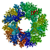
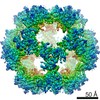
| Title | Atomic Structure of the E2 Inner Core of Human Pyruvate Dehydrogenase Complex | |||||||||||||||||||||||||||
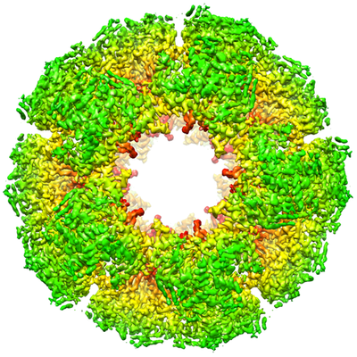
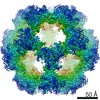
 Map data Map data | 3D reconstruction of PDC tE2 after particle polishing | |||||||||||||||||||||||||||
 Sample Sample |
| |||||||||||||||||||||||||||
 Keywords Keywords | Pyruvate Dehydrogenase Complex / E2 / Inner Core / OXIDOREDUCTASE | |||||||||||||||||||||||||||
| Function / homology |  Function and homology information Function and homology informationPDH complex synthesizes acetyl-CoA from PYR / dihydrolipoyllysine-residue acetyltransferase / dihydrolipoyllysine-residue acetyltransferase activity / pyruvate decarboxylation to acetyl-CoA / Regulation of pyruvate dehydrogenase (PDH) complex / Protein lipoylation / pyruvate catabolic process / pyruvate dehydrogenase complex / Signaling by Retinoic Acid / sperm head-tail coupling apparatus ...PDH complex synthesizes acetyl-CoA from PYR / dihydrolipoyllysine-residue acetyltransferase / dihydrolipoyllysine-residue acetyltransferase activity / pyruvate decarboxylation to acetyl-CoA / Regulation of pyruvate dehydrogenase (PDH) complex / Protein lipoylation / pyruvate catabolic process / pyruvate dehydrogenase complex / Signaling by Retinoic Acid / sperm head-tail coupling apparatus / tricarboxylic acid cycle / glucose metabolic process / mitochondrial matrix / mitochondrion / identical protein binding Similarity search - Function | |||||||||||||||||||||||||||
| Biological species |  Homo sapiens (human) Homo sapiens (human) | |||||||||||||||||||||||||||
| Method | single particle reconstruction / cryo EM / Resolution: 3.1 Å | |||||||||||||||||||||||||||
 Authors Authors | Jiang J / Baiesc FL | |||||||||||||||||||||||||||
| Funding support |  United States, 8 items United States, 8 items
| |||||||||||||||||||||||||||
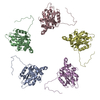
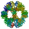
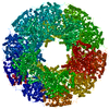
 Citation Citation | Journal: Biochemistry / Year: 2018 Title: Atomic Structure of the E2 Inner Core of Human Pyruvate Dehydrogenase Complex. Authors: Jiansen Jiang / Flavius L Baiesc / Yasuaki Hiromasa / Xuekui Yu / Wong Hoi Hui / Xinghong Dai / Thomas E Roche / Z Hong Zhou /  Abstract: Pyruvate dehydrogenase complex (PDC) is a large multienzyme complex that catalyzes the irreversible conversion of pyruvate to acetyl-coenzyme A with reduction of NAD. Distinctive from PDCs in lower ...Pyruvate dehydrogenase complex (PDC) is a large multienzyme complex that catalyzes the irreversible conversion of pyruvate to acetyl-coenzyme A with reduction of NAD. Distinctive from PDCs in lower forms of life, in mammalian PDC, dihydrolipoyl acetyltransferase (E2; E2p in PDC) and dihydrolipoamide dehydrogenase binding protein (E3BP) combine to form a complex that plays a central role in the organization, regulation, and integration of catalytic reactions of PDC. However, the atomic structure and organization of the mammalian E2p/E3BP heterocomplex are unknown. Here, we report the structure of the recombinant dodecahedral core formed by the C-terminal inner-core/catalytic (IC) domain of human E2p determined at 3.1 Å resolution by cryo electron microscopy (cryoEM). The structure of the N-terminal fragment and four other surface areas of the human E2p IC domain exhibit significant differences from those of the other E2 crystal structures, which may have implications for the integration of E3BP in mammals. This structure also allowed us to obtain a homology model for the highly homologous IC domain of E3BP. Analysis of the interactions of human E2p or E3BP with their adjacent IC domains in the dodecahedron provides new insights into the organization of the E2p/E3BP heterocomplex and suggests a potential contribution by E3BP to catalysis in mammalian PDC. | |||||||||||||||||||||||||||
| History |
|
- Structure visualization
| Movie |
Movie viewer |
|---|---|
| Structure viewer | EM map: SurfViewMolmilJmol/JSmol |
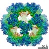
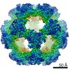
| Supplemental images |
- Downloads & links
-EMDB archive
| Map data | emd_7610.map.gz | 59.5 MB | EMDB map data format | |
|---|---|---|---|---|
| Header (meta data) | emd-7610-v30.xmlemd-7610.xml | 12.1 KB 12.1 KB | Display Display | EMDB header |
| Images |  emd_7610.png emd_7610.png | 269.9 KB | ||
| Filedesc metadata | emd-7610.cif.gz | 5.3 KB | ||
| Archive directory |  http://ftp.pdbj.org/pub/emdb/structures/EMD-7610ftp://ftp.pdbj.org/pub/emdb/structures/EMD-7610 http://ftp.pdbj.org/pub/emdb/structures/EMD-7610ftp://ftp.pdbj.org/pub/emdb/structures/EMD-7610 | HTTPS FTP |
-Related structure data
| Related structure data | 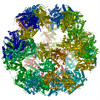 6ct0MC M: atomic model generated by this map C: citing same article ( |
|---|---|
| Similar structure data |
-Links
| EMDB pages | EMDB (EBI/PDBe) / EMDataResource |
|---|---|
| Related items in Molecule of the Month |


-Map
| File | Download / File: emd_7610.map.gz / Format: CCP4 / Size: 64 MB / Type: IMAGE STORED AS FLOATING POINT NUMBER (4 BYTES) | ||||||||||||||||||||||||||||||||||||||||||||||||||||||||||||||||||||
|---|---|---|---|---|---|---|---|---|---|---|---|---|---|---|---|---|---|---|---|---|---|---|---|---|---|---|---|---|---|---|---|---|---|---|---|---|---|---|---|---|---|---|---|---|---|---|---|---|---|---|---|---|---|---|---|---|---|---|---|---|---|---|---|---|---|---|---|---|---|
| Annotation | 3D reconstruction of PDC tE2 after particle polishing | ||||||||||||||||||||||||||||||||||||||||||||||||||||||||||||||||||||
| Projections & slices | Image control
Images are generated by Spider. | ||||||||||||||||||||||||||||||||||||||||||||||||||||||||||||||||||||
| Voxel size | X=Y=Z: 1.02 Å | ||||||||||||||||||||||||||||||||||||||||||||||||||||||||||||||||||||
| Density |
| ||||||||||||||||||||||||||||||||||||||||||||||||||||||||||||||||||||
| Symmetry | Space group: 1 | ||||||||||||||||||||||||||||||||||||||||||||||||||||||||||||||||||||
| Details | EMDB XML:
CCP4 map header:
| ||||||||||||||||||||||||||||||||||||||||||||||||||||||||||||||||||||
 Z (Sec.)
 Y (Row.)
 X (Col.)


-Supplemental data
- Sample components
-Entire : E2 Inner Core of Human Pyruvate Dehydrogenase Complex
| Entire | Name: E2 Inner Core of Human Pyruvate Dehydrogenase Complex |
|---|---|
| Components |
|
-Supramolecule #1: E2 Inner Core of Human Pyruvate Dehydrogenase Complex
| Supramolecule | Name: E2 Inner Core of Human Pyruvate Dehydrogenase Complex / type: complex / ID: 1 / Parent: 0 / Macromolecule list: all |
|---|---|
| Source (natural) | Organism: Homo sapiens (human) |
-Macromolecule #1: Dihydrolipoyllysine-residue acetyltransferase component of pyruva...
| Macromolecule | Name: Dihydrolipoyllysine-residue acetyltransferase component of pyruvate dehydrogenase complex, mitochondrial type: protein_or_peptide / ID: 1 / Number of copies: 1 / Enantiomer: LEVO / EC number: dihydrolipoyllysine-residue acetyltransferase |
|---|---|
| Source (natural) | Organism: Homo sapiens (human) |
| Molecular weight | Theoretical: 69.069398 KDa |
| Recombinant expression | Organism:  |
| Sequence | String: MWRVCARRAQ NVAPWAGLEA RWTALQEVPG TPRVTSRSGP APARRNSVTT GYGGVRALCG WTPSSGATPR NRLLLQLLGS PGRRYYSLP PHQKVPLPSL SPTMQAGTIA RWEKKEGDKI NEGDLIAEVE TDKATVGFES LEECYMAKIL VAEGTRDVPI G AIICITVG ...String: MWRVCARRAQ NVAPWAGLEA RWTALQEVPG TPRVTSRSGP APARRNSVTT GYGGVRALCG WTPSSGATPR NRLLLQLLGS PGRRYYSLP PHQKVPLPSL SPTMQAGTIA RWEKKEGDKI NEGDLIAEVE TDKATVGFES LEECYMAKIL VAEGTRDVPI G AIICITVG KPEDIEAFKN YTLDSSAAPT PQAAPAPTPA ATASPPTPSA QAPGSSYPPH MQVLLPALSP TMTMGTVQRW EK KVGEKLS EGDLLAEIET DKATIGFEVQ EEGYLAKILV PEGTRDVPLG TPLCIIVEKE ADISAFADYR PTEVTDLKPQ VPP PTPPPV AAVPPTPQPL APTPSAPCPA TPAGPKGRVF VSPLAKKLAV EKGIDLTQVK GTGPDGRITK KDIDSFVPSK VAPA PAAVV PPTGPGMAPV PTGVFTDIPI SNIRRVIAQR LMQSKQTIPH YYLSIDVNMG EVLLVRKELN KILEGRSKIS VNDFI IKAS ALACLKVPEA NSSWMDTVIR QNHVVDVSVA VSTPAGLITP IVFNAHIKGV ETIANDVVSL ATKAREGKLQ PHEFQG GTF TISNLGMFGI KNFSAIINPP QACILAIGAS EDKLVPADNE KGFDVASMMS VTLSCDHRVV DGAVGAQWLA EFRKYLE KP ITMLL UniProtKB: Dihydrolipoyllysine-residue acetyltransferase component of pyruvate dehydrogenase complex, mitochondrial |
-Experimental details
-Structure determination
| Method | cryo EM |
|---|---|
 Processing Processing | single particle reconstruction |
| Aggregation state | particle |
-Sample preparation
| Buffer | pH: 7.2 |
|---|---|
| Vitrification | Cryogen name: ETHANE |
- Electron microscopy
| Microscope | FEI TITAN KRIOS |
|---|---|
| Image recording | Film or detector model: GATAN K2 SUMMIT (4k x 4k) / Detector mode: COUNTING / Average exposure time: 8.0 sec. / Average electron dose: 38.0 e/Å2 |
| Electron beam | Acceleration voltage: 300 kV / Electron source:  FIELD EMISSION GUN FIELD EMISSION GUN |
| Electron optics | Illumination mode: FLOOD BEAM / Imaging mode: BRIGHT FIELD |
| Experimental equipment |  Model: Titan Krios / Image courtesy: FEI Company |
-Image processing
| Startup model | Type of model: NONE |
|---|---|
| Final reconstruction | Resolution.type: BY AUTHOR / Resolution: 3.1 Å / Resolution method: FSC 0.143 CUT-OFF / Software - Name: RELION / Number images used: 49374 |
| Initial angle assignment | Type: NOT APPLICABLE / Software - Name: RELION |
| Final angle assignment | Type: NOT APPLICABLE / Software - Name: RELION |
