 Movie
Movie Controller

[English] 日本語
 Yorodumi
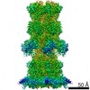
- EMDB-10912: Cryo-EM structure of T7 bacteriophage DNA translocation gp15-gp16... -
+ Open data

- Basic information
| Entry | Database: EMDB / ID: EMD-10912 | ||||||||||||
|---|---|---|---|---|---|---|---|---|---|---|---|---|---|
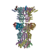
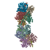
| Title | Cryo-EM structure of T7 bacteriophage DNA translocation gp15-gp16 core complex intermediate assembly | ||||||||||||
 Map data Map data | t7 tail complex | ||||||||||||
 Sample Sample |
| ||||||||||||
 Keywords Keywords | VIRAL COMPLEX / DNA TRANSLOCATION / VIRAL INFECTION / TRANS-GLYCOSYLASE ACTIVITY / CHAPERONE / PERIPLASMIC SPACE COMPLEX / VIRAL PROTEIN | ||||||||||||
| Function / homology |  Function and homology information Function and homology informationsymbiont genome ejection through host cell envelope / host cell periplasmic space / : / symbiont entry into host cell via disruption of host cell wall peptidoglycan / peptidoglycan lytic transglycosylase activity / symbiont genome ejection through host cell envelope, short tail mechanism / peptidoglycan metabolic process / symbiont entry into host cell via disruption of host cell envelope / symbiont entry into host / virion component ...symbiont genome ejection through host cell envelope / host cell periplasmic space / : / symbiont entry into host cell via disruption of host cell wall peptidoglycan / peptidoglycan lytic transglycosylase activity / symbiont genome ejection through host cell envelope, short tail mechanism / peptidoglycan metabolic process / symbiont entry into host cell via disruption of host cell envelope / symbiont entry into host / virion component / killing of cells of another organism / defense response to bacterium / hydrolase activity / host cell plasma membrane / membrane Similarity search - Function | ||||||||||||
| Biological species |   Escherichia phage T7 (virus) Escherichia phage T7 (virus) | ||||||||||||
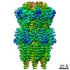
| Method | single particle reconstruction / cryo EM / Resolution: 3.0 Å | ||||||||||||
 Authors Authors | Perez-Ruiz M / Pulido-Cid M | ||||||||||||
| Funding support |  Spain, 3 items Spain, 3 items
| ||||||||||||
 Citation Citation | Journal: Proc Natl Acad Sci U S A / Year: 2021 Title: Assisted assembly of bacteriophage T7 core components for genome translocation across the bacterial envelope. Authors: Mar Pérez-Ruiz / Mar Pulido-Cid / Juan Román Luque-Ortega / José María Valpuesta / Ana Cuervo / José L Carrascosa / Abstract: In most bacteriophages, genome transport across bacterial envelopes is carried out by the tail machinery. In viruses of the family, in which the tail is not long enough to traverse the bacterial ...In most bacteriophages, genome transport across bacterial envelopes is carried out by the tail machinery. In viruses of the family, in which the tail is not long enough to traverse the bacterial wall, it has been postulated that viral core proteins assembled inside the viral head are translocated and reassembled into a tube within the periplasm that extends the tail channel. Bacteriophage T7 infects , and despite extensive studies, the precise mechanism by which its genome is translocated remains unknown. Using cryo-electron microscopy, we have resolved the structure of two different assemblies of the T7 DNA translocation complex composed of the core proteins gp15 and gp16. Gp15 alone forms a partially folded hexamer, which is further assembled upon interaction with gp16 into a tubular structure, forming a channel that could allow DNA passage. The structure of the gp15-gp16 complex also shows the location within gp16 of a canonical transglycosylase motif involved in the degradation of the bacterial peptidoglycan layer. This complex docks well in the tail extension structure found in the periplasm of T7-infected bacteria and matches the sixfold symmetry of the phage tail. In such cases, gp15 and gp16 that are initially present in the T7 capsid eightfold-symmetric core would change their oligomeric state upon reassembly in the periplasm. Altogether, these results allow us to propose a model for the assembly of the core translocation complex in the periplasm, which furthers understanding of the molecular mechanism involved in the release of T7 viral DNA into the bacterial cytoplasm. | ||||||||||||
| History |
|
- Structure visualization
| Movie |
Movie viewer |
|---|---|
| Structure viewer | EM map: SurfViewMolmilJmol/JSmol |
| Supplemental images |

- Downloads & links
-EMDB archive
| Map data | emd_10912.map.gz | 111.5 MB | EMDB map data format | |
|---|---|---|---|---|
| Header (meta data) | emd-10912-v30.xmlemd-10912.xml | 17.1 KB 17.1 KB | Display Display | EMDB header |
| FSC (resolution estimation) | emd_10912_fsc.xml | 11.4 KB | Display | FSC data file |
| Images | 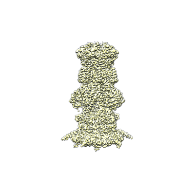 emd_10912.png emd_10912.png | 70.3 KB | ||
| Filedesc metadata | emd-10912.cif.gz | 7.1 KB | ||
| Archive directory |  http://ftp.pdbj.org/pub/emdb/structures/EMD-10912ftp://ftp.pdbj.org/pub/emdb/structures/EMD-10912 http://ftp.pdbj.org/pub/emdb/structures/EMD-10912ftp://ftp.pdbj.org/pub/emdb/structures/EMD-10912 | HTTPS FTP |
-Related structure data
| Related structure data | 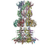 6yt5MC 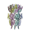 6yszC C: citing same article ( M: atomic model generated by this map |
|---|---|
| Similar structure data |


-Links
| EMDB pages | EMDB (EBI/PDBe) / EMDataResource |
|---|
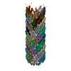
-Map
| File | Download / File: emd_10912.map.gz / Format: CCP4 / Size: 125 MB / Type: IMAGE STORED AS FLOATING POINT NUMBER (4 BYTES) | ||||||||||||||||||||||||||||||||||||||||||||||||||||||||||||
|---|---|---|---|---|---|---|---|---|---|---|---|---|---|---|---|---|---|---|---|---|---|---|---|---|---|---|---|---|---|---|---|---|---|---|---|---|---|---|---|---|---|---|---|---|---|---|---|---|---|---|---|---|---|---|---|---|---|---|---|---|---|
| Annotation | t7 tail complex | ||||||||||||||||||||||||||||||||||||||||||||||||||||||||||||
| Projections & slices | Image control
Images are generated by Spider. | ||||||||||||||||||||||||||||||||||||||||||||||||||||||||||||
| Voxel size | X=Y=Z: 1.047 Å | ||||||||||||||||||||||||||||||||||||||||||||||||||||||||||||
| Density |
| ||||||||||||||||||||||||||||||||||||||||||||||||||||||||||||
| Symmetry | Space group: 1 | ||||||||||||||||||||||||||||||||||||||||||||||||||||||||||||
| Details | EMDB XML:
CCP4 map header:
| ||||||||||||||||||||||||||||||||||||||||||||||||||||||||||||
 Z (Sec.)
 Y (Row.)
 X (Col.)


-Supplemental data
- Sample components
-Entire : gp15-gp16 core complex
| Entire | Name: gp15-gp16 core complex |
|---|---|
| Components |
|
-Supramolecule #1: gp15-gp16 core complex
| Supramolecule | Name: gp15-gp16 core complex / type: complex / ID: 1 / Parent: 0 / Macromolecule list: all |
|---|---|
| Source (natural) | Organism: Escherichia phage T7 (virus) |
| Molecular weight | Theoretical: 1.4 MDa |
-Macromolecule #1: Internal virion protein gp15
| Macromolecule | Name: Internal virion protein gp15 / type: protein_or_peptide / ID: 1 / Number of copies: 6 / Enantiomer: LEVO |
|---|---|
| Source (natural) | Organism: Escherichia phage T7 (virus) |
| Molecular weight | Theoretical: 88.377219 KDa |
| Recombinant expression | Organism:  |
| Sequence | String: MRGSHHHHHH GMASMTGGQQ MGRDLYDDDD KDPSSMSKIE SALQAAQPGL SRLRGGAGGM GYRAATTQAE QPRSSLLDTI GRFAKAGAD MYTAKEQRAR DLADERSNEI IRKLTPEQRR EALNNGTLLY QDDPYAMEAL RVKTGRNAAY LVDDDVMQKI K EGVFRTRE ...String: MRGSHHHHHH GMASMTGGQQ MGRDLYDDDD KDPSSMSKIE SALQAAQPGL SRLRGGAGGM GYRAATTQAE QPRSSLLDTI GRFAKAGAD MYTAKEQRAR DLADERSNEI IRKLTPEQRR EALNNGTLLY QDDPYAMEAL RVKTGRNAAY LVDDDVMQKI K EGVFRTRE EMEEYRHSRL QEGAKVYAEQ FGIDPEDVDY QRGFNGDITE RNISLYGAHD NFLSQQAQKG AIMNSRVELN GV LQDPDML RRPDSADFFE KYIDNGLVTG AIPSDAQATQ LISQAFSDAS SRAGGADFLM RVGDKKVTLN GATTTYRELI GEE QWNALM VTAQRSQFET DAKLNEQYRL KINSALNQED PRTAWEMLQG IKAELDKVQP DEQMTPQREW LISAQEQVQN QMNA WTKAQ AKALDDSMKS MNKLDVIDKQ FQKRINGEWV STDFKDMPVN ENTGEFKHSD MVNYANKKLA EIDSMDIPDG AKDAM KLKY LQADSKDGAF RTAIGTMVTD AGQEWSAAVI NGKLPERTPA MDALRRIRNA DPQLIAALYP DQAELFLTMD MMDKQG IDP QVILDADRLT VKRSKEQRFE DDKAFESALN ASKAPEIARM PASLRESARK IYDSVKYRSG NESMAMEQMT KFLKEST YT FTGDDVDGDT VGVIPKNMMQ VNSDPKSWEQ GRDILEEARK GIIASNPWIT NKQLTMYSQG DSIYLMDTTG QVRVRYDK E LLSKVWSENQ KKLEEKAREK ALADVNKRAP IVAATKAREA AAKRVREKRK QTPKFIYGRK E UniProtKB: Internal virion protein gp15 |
-Macromolecule #2: Peptidoglycan transglycosylase gp16
| Macromolecule | Name: Peptidoglycan transglycosylase gp16 / type: protein_or_peptide / ID: 2 / Number of copies: 6 / Enantiomer: LEVO EC number: Lyases; Carbon-oxygen lyases; Acting on polysaccharides |
|---|---|
| Source (natural) | Organism: Escherichia phage T7 (virus) |
| Molecular weight | Theoretical: 146.693094 KDa |
| Recombinant expression | Organism: |
| Sequence | String: MGHHHHHHHH HHSSGHIEGR HMMDKYDKNV PSDYDGLFQK AADANGVSYD LLRKVAWTES RFVPTAKSKT GPLGMMQFTK ATAKALGLR VTDGPDDDRL NPELAINAAA KQLAGLVGKF DGDELKAALA YNQGEGRLGN PQLEAYSKGD FASISEEGRN Y MRNLLDVA ...String: MGHHHHHHHH HHSSGHIEGR HMMDKYDKNV PSDYDGLFQK AADANGVSYD LLRKVAWTES RFVPTAKSKT GPLGMMQFTK ATAKALGLR VTDGPDDDRL NPELAINAAA KQLAGLVGKF DGDELKAALA YNQGEGRLGN PQLEAYSKGD FASISEEGRN Y MRNLLDVA KSPMAGQLET FGGITPKGKG IPAEVGLAGI GHKQKVTQEL PESTSFDVKG IEQEATAKPF AKDFWETHGE TL DEYNSRS TFFGFKNAAE AELSNSVAGM AFRAGRLDNG FDVFKDTITP TRWNSHIWTP EELEKIRTEV KNPAYINVVT GGS PENLDD LIKLANENFE NDSRAAEAGL GAKLSAGIIG AGVDPLSYVP MVGVTGKGFK LINKALVVGA ESAALNVASE GLRT SVAGG DADYAGAALG GFVFGAGMSA ISDAVAAGLK RSKPEAEFDN EFIGPMMRLE ARETARNANS ADLSRMNTEN MKFEG EHNG VPYEDLPTER GAVVLHDGSV LSASNPINPK TLKEFSEVDP EKAARGIKLA GFTEIGLKTL GSDDADIRRV AIDLVR SPT GMQSGASGKF GATASDIHER LHGTDQRTYN DLYKAMSDAM KDPEFSTGGA KMSREETRYT IYRRAALAIE RPELQKA LT PSERIVMDII KRHFDTKREL MENPAIFGNT KAVSIFPESR HKGTYVPHVY DRHAKALMIQ RYGAEGLQEG IARSWMNS Y VSRPEVKARV DEMLKELHGV KEVTPEMVEK YAMDKAYGIS HSDQFTNSSI IEENIEGLVG IENNSFLEAR NLFDSDLSI TMPDGQQFSV NDLRDFDMFR IMPAYDRRVN GDIAIMGSTG KTTKELKDEI LALKAKAEGD GKKTGEVHAL MDTVKILTGR ARRNQDTVW ETSLRAINDL GFFAKNAYMG AQNITEIAGM IVTGNVRALG HGIPILRDTL YKSKPVSAKE LKELHASLFG K EVDQLIRP KRADIVQRLR EATDTGPAVA NIVGTLKYST QELAARSPWT KLLNGTTNYL LDAARQGMLG DVISATLTGK TT RWEKEGF LRGASVTPEQ MAGIKSLIKE HMVRGEDGKF TVKDKQAFSM DPRAMDLWRL ADKVADEAML RPHKVSLQDS HAF GALGKM VMQFKSFTIK SLNSKFLRTF YDGYKNNRAI DAALSIITSM GLAGGFYAMA AHVKAYALPK EKRKEYLERA LDPT MIAHA ALSRSSQLGA PLAMVDLVGG VLGFESSKMA RSTILPKDTV KERDPNKPYT SREVMGAMGS NLLEQMPSAG FVANV GATL MNAAGVVNSP NKATEQDFMT GLMNSTKELV PNDPLTQQLV LKIYEANGVN LRERRK UniProtKB: Peptidoglycan transglycosylase gp16 |
-Experimental details
-Structure determination
| Method | cryo EM |
|---|---|
 Processing Processing | single particle reconstruction |
| Aggregation state | particle |
-Sample preparation
| Concentration | 1.5 mg/mL | ||||||||
|---|---|---|---|---|---|---|---|---|---|
| Buffer | pH: 8 Component:
| ||||||||
| Grid | Model: Quantifoil R2/2 / Pretreatment - Type: GLOW DISCHARGE / Pretreatment - Time: 60 sec. / Pretreatment - Atmosphere: AIR | ||||||||
| Vitrification | Cryogen name: ETHANE / Chamber humidity: 95 % / Chamber temperature: 293.15 K / Instrument: FEI VITROBOT MARK IV |
- Electron microscopy
| Microscope | FEI TITAN KRIOS |
|---|---|
| Image recording | Film or detector model: GATAN K2 SUMMIT (4k x 4k) / Detector mode: COUNTING / Number grids imaged: 2 / Number real images: 5506 / Average exposure time: 8.0 sec. / Average electron dose: 40.8 e/Å2 |
| Electron beam | Acceleration voltage: 300 kV / Electron source:  FIELD EMISSION GUN FIELD EMISSION GUN |
| Electron optics | Calibrated magnification: 47619 / Illumination mode: FLOOD BEAM / Imaging mode: BRIGHT FIELD |
| Sample stage | Specimen holder model: FEI TITAN KRIOS AUTOGRID HOLDER / Cooling holder cryogen: NITROGEN |
| Experimental equipment |  Model: Titan Krios / Image courtesy: FEI Company |
+Image processing

-Atomic model buiding 1
| Initial model |
| ||||||
|---|---|---|---|---|---|---|---|
| Refinement | Space: REAL / Protocol: AB INITIO MODEL | ||||||
| Output model | PDB-6yt5: |


