 Movie
Movie Controller

[English] 日本語
 Yorodumi
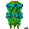
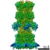
- PDB-6yt5: Cryo-EM structure of T7 bacteriophage DNA translocation gp15-gp16... -
+ Open data

- Basic information
| Entry | Database: PDB / ID: 6yt5 | ||||||||||||
|---|---|---|---|---|---|---|---|---|---|---|---|---|---|
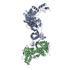
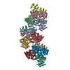
| Title | Cryo-EM structure of T7 bacteriophage DNA translocation gp15-gp16 core complex intermediate assembly | ||||||||||||
 Components Components |
| ||||||||||||
 Keywords Keywords | VIRAL PROTEIN / VIRAL COMPLEX / DNA TRANSLOCATION / VIRAL INFECTION / TRANS-GLYCOSYLASE ACTIVITY / CHAPERONE / PERIPLASMIC SPACE COMPLEX | ||||||||||||
| Function / homology |  Function and homology information Function and homology informationsymbiont genome ejection through host cell envelope / host cell periplasmic space / : / symbiont entry into host cell via disruption of host cell wall peptidoglycan / peptidoglycan lytic transglycosylase activity / symbiont genome ejection through host cell envelope, short tail mechanism / peptidoglycan metabolic process / symbiont entry into host cell via disruption of host cell envelope / symbiont entry into host / virion component ...symbiont genome ejection through host cell envelope / host cell periplasmic space / : / symbiont entry into host cell via disruption of host cell wall peptidoglycan / peptidoglycan lytic transglycosylase activity / symbiont genome ejection through host cell envelope, short tail mechanism / peptidoglycan metabolic process / symbiont entry into host cell via disruption of host cell envelope / symbiont entry into host / virion component / killing of cells of another organism / defense response to bacterium / hydrolase activity / host cell plasma membrane / membrane Similarity search - Function | ||||||||||||
| Biological species |   Escherichia phage T7 (virus) Escherichia phage T7 (virus) | ||||||||||||
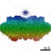
| Method | ELECTRON MICROSCOPY / single particle reconstruction / cryo EM / Resolution: 3 Å | ||||||||||||
 Authors Authors | Perez-Ruiz, M. / Pulido-Cid, M. / Luque-Ortega, J.R. / Cuervo, A. / Carrascosa, J.L. | ||||||||||||
| Funding support |  Spain, 3items Spain, 3items
| ||||||||||||
 Citation Citation | Journal: Proc Natl Acad Sci U S A / Year: 2021 Title: Assisted assembly of bacteriophage T7 core components for genome translocation across the bacterial envelope. Authors: Mar Pérez-Ruiz / Mar Pulido-Cid / Juan Román Luque-Ortega / José María Valpuesta / Ana Cuervo / José L Carrascosa / Abstract: In most bacteriophages, genome transport across bacterial envelopes is carried out by the tail machinery. In viruses of the family, in which the tail is not long enough to traverse the bacterial ...In most bacteriophages, genome transport across bacterial envelopes is carried out by the tail machinery. In viruses of the family, in which the tail is not long enough to traverse the bacterial wall, it has been postulated that viral core proteins assembled inside the viral head are translocated and reassembled into a tube within the periplasm that extends the tail channel. Bacteriophage T7 infects , and despite extensive studies, the precise mechanism by which its genome is translocated remains unknown. Using cryo-electron microscopy, we have resolved the structure of two different assemblies of the T7 DNA translocation complex composed of the core proteins gp15 and gp16. Gp15 alone forms a partially folded hexamer, which is further assembled upon interaction with gp16 into a tubular structure, forming a channel that could allow DNA passage. The structure of the gp15-gp16 complex also shows the location within gp16 of a canonical transglycosylase motif involved in the degradation of the bacterial peptidoglycan layer. This complex docks well in the tail extension structure found in the periplasm of T7-infected bacteria and matches the sixfold symmetry of the phage tail. In such cases, gp15 and gp16 that are initially present in the T7 capsid eightfold-symmetric core would change their oligomeric state upon reassembly in the periplasm. Altogether, these results allow us to propose a model for the assembly of the core translocation complex in the periplasm, which furthers understanding of the molecular mechanism involved in the release of T7 viral DNA into the bacterial cytoplasm. | ||||||||||||
| History |
|
- Structure visualization
| Movie |
Movie viewer |
|---|---|
| Structure viewer | Molecule: MolmilJmol/JSmol |
- Downloads & links
-Download
| PDBx/mmCIF format | 6yt5.cif.gz | 984.1 KB | Display | PDBx/mmCIF format |
|---|---|---|---|---|
| PDB format | pdb6yt5.ent.gz | 726.9 KB | Display | PDB format |
| PDBx/mmJSON format | 6yt5.json.gz | Tree view | PDBx/mmJSON format | |
| Others |  Other downloads Other downloads |
-Validation report
| Arichive directory | https://data.pdbj.org/pub/pdb/validation_reports/yt/6yt5ftp://data.pdbj.org/pub/pdb/validation_reports/yt/6yt5 | HTTPS FTP |
|---|
-Related structure data
| Related structure data | 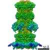 10912MC 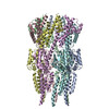 6yszC C: citing same article ( M: map data used to model this data |
|---|---|
| Similar structure data |

-Links
 PDBj
PDBj
- Assembly
| Deposited unit | 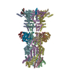
|
|---|---|
| 1 |
|
-Components
| #1: Protein | Mass: 88377.219 Da / Num. of mol.: 6 Source method: isolated from a genetically manipulated source Source: (gene. exp.) Escherichia phage T7 (virus) / Gene: 15 / Production host:  #2: Protein | Mass: 146693.094 Da / Num. of mol.: 6 Source method: isolated from a genetically manipulated source Source: (gene. exp.) Escherichia phage T7 (virus) / Gene: 16 / Production host: References: UniProt: P03726, Lyases; Carbon-oxygen lyases; Acting on polysaccharides |
|---|
-Experimental details
-Experiment
| Experiment | Method: ELECTRON MICROSCOPY |
|---|---|
| EM experiment | Aggregation state: PARTICLE / 3D reconstruction method: single particle reconstruction |
- Sample preparation
| Component | Name: gp15-gp16 core complex / Type: COMPLEX / Entity ID: all / Source: RECOMBINANT | ||||||||||||||||
|---|---|---|---|---|---|---|---|---|---|---|---|---|---|---|---|---|---|
| Molecular weight | Value: 1.4 MDa / Experimental value: YES | ||||||||||||||||
| Source (natural) | Organism: Escherichia phage T7 (virus) | ||||||||||||||||
| Source (recombinant) | Organism: | ||||||||||||||||
| Buffer solution | pH: 8 | ||||||||||||||||
| Buffer component |
| ||||||||||||||||
| Specimen | Conc.: 1.5 mg/ml / Embedding applied: NO / Shadowing applied: NO / Staining applied: NO / Vitrification applied: YES | ||||||||||||||||
| Specimen support | Grid type: Quantifoil R2/2 | ||||||||||||||||
| Vitrification | Instrument: FEI VITROBOT MARK IV / Cryogen name: ETHANE / Humidity: 95 % / Chamber temperature: 293.15 K |
- Electron microscopy imaging
| Experimental equipment |  Model: Titan Krios / Image courtesy: FEI Company |
|---|---|
| Microscopy | Model: FEI TITAN KRIOS |
| Electron gun | Electron source:  FIELD EMISSION GUN / Accelerating voltage: 300 kV / Illumination mode: FLOOD BEAM FIELD EMISSION GUN / Accelerating voltage: 300 kV / Illumination mode: FLOOD BEAM |
| Electron lens | Mode: BRIGHT FIELD / Calibrated magnification: 47619 X / Alignment procedure: COMA FREE |
| Specimen holder | Cryogen: NITROGEN / Specimen holder model: FEI TITAN KRIOS AUTOGRID HOLDER |
| Image recording | Average exposure time: 8 sec. / Electron dose: 40.8 e/Å2 / Detector mode: COUNTING / Film or detector model: GATAN K2 SUMMIT (4k x 4k) / Num. of grids imaged: 2 / Num. of real images: 5506 |
| Image scans | Movie frames/image: 32 |
- Processing
| EM software |
| ||||||||||||||||||||||||||||||||||||||||||||
|---|---|---|---|---|---|---|---|---|---|---|---|---|---|---|---|---|---|---|---|---|---|---|---|---|---|---|---|---|---|---|---|---|---|---|---|---|---|---|---|---|---|---|---|---|---|
| CTF correction | Type: PHASE FLIPPING AND AMPLITUDE CORRECTION | ||||||||||||||||||||||||||||||||||||||||||||
| Symmetry | Point symmetry: C6 (6 fold cyclic) | ||||||||||||||||||||||||||||||||||||||||||||
| 3D reconstruction | Resolution: 3 Å / Resolution method: FSC 0.143 CUT-OFF / Num. of particles: 72882 / Algorithm: FOURIER SPACE / Num. of class averages: 1 / Symmetry type: POINT | ||||||||||||||||||||||||||||||||||||||||||||
| Atomic model building | Protocol: AB INITIO MODEL / Space: REAL | ||||||||||||||||||||||||||||||||||||||||||||
| Atomic model building | 3D fitting-ID: 1 / Pdb chain-ID: A / Source name: PDB / Type: experimental model
|


