 Movie
Movie Controller


+ Open data

- Basic information
| Entry | Database: EMDB / ID: EMD-22680 | |||||||||
|---|---|---|---|---|---|---|---|---|---|---|
| Title | Structure of T7 DNA ejectosome periplasmic tunnel | |||||||||
 Map data Map data | Post-process map from Relion-3.0 | |||||||||
 Sample Sample |
| |||||||||
 Keywords Keywords | T7 / ejectosome / ejection protein / genome-delivery / podoviridae / VIRAL PROTEIN | |||||||||
| Function / homology |  Function and homology information Function and homology informationsymbiont genome ejection through host cell envelope / host cell periplasmic space / : / symbiont entry into host cell via disruption of host cell wall peptidoglycan / peptidoglycan lytic transglycosylase activity / symbiont genome ejection through host cell envelope, short tail mechanism / peptidoglycan metabolic process / symbiont entry into host cell via disruption of host cell envelope / symbiont entry into host / virion component ...symbiont genome ejection through host cell envelope / host cell periplasmic space / : / symbiont entry into host cell via disruption of host cell wall peptidoglycan / peptidoglycan lytic transglycosylase activity / symbiont genome ejection through host cell envelope, short tail mechanism / peptidoglycan metabolic process / symbiont entry into host cell via disruption of host cell envelope / symbiont entry into host / virion component / killing of cells of another organism / defense response to bacterium / hydrolase activity / host cell plasma membrane / membrane Similarity search - Function | |||||||||
| Biological species |   Escherichia phage T7 (virus) Escherichia phage T7 (virus) | |||||||||
| Method | single particle reconstruction / cryo EM / Resolution: 2.7 Å | |||||||||
 Authors Authors | Swanson N / Cingolani G | |||||||||
| Funding support |  United States, 1 items United States, 1 items
| |||||||||
 Citation Citation | Journal: Mol Cell / Year: 2021 Title: Cryo-EM structure of the periplasmic tunnel of T7 DNA-ejectosome at 2.7 Å resolution. Authors: Nicholas A Swanson / Ravi K Lokareddy / Fenglin Li / Chun-Feng David Hou / Sebastian Leptihn / Mikhail Pavlenok / Michael Niederweis / Ruth A Pumroy / Vera Y Moiseenkova-Bell / Gino Cingolani /  Abstract: Hershey and Chase used bacteriophage T2 genome delivery inside Escherichia coli to demonstrate that DNA, not protein, is the genetic material. Seventy years later, our understanding of viral genome ...Hershey and Chase used bacteriophage T2 genome delivery inside Escherichia coli to demonstrate that DNA, not protein, is the genetic material. Seventy years later, our understanding of viral genome delivery in prokaryotes remains limited, especially for short-tailed phages of the Podoviridae family. These viruses expel mysterious ejection proteins found inside the capsid to form a DNA-ejectosome for genome delivery into bacteria. Here, we reconstitute the phage T7 DNA-ejectosome components gp14, gp15, and gp16 and solve the periplasmic tunnel structure at 2.7 Å resolution. We find that gp14 forms an outer membrane pore, gp15 assembles into a 210 Å hexameric DNA tube spanning the host periplasm, and gp16 extends into the host cytoplasm forming a ∼4,200 residue hub. Gp16 promotes gp15 oligomerization, coordinating peptidoglycan hydrolysis, DNA binding, and lipid insertion. The reconstituted gp15:gp16 complex lacks channel-forming activity, suggesting that the pore for DNA passage forms only transiently during genome ejection. | |||||||||
| History |
|
- Structure visualization
| Movie |
Movie viewer |
|---|---|
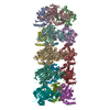
| Structure viewer | EM map: SurfViewMolmilJmol/JSmol |
| Supplemental images |

- Downloads & links
-EMDB archive
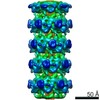
| Map data | emd_22680.map.gz | 156.1 MB | EMDB map data format | |
|---|---|---|---|---|
| Header (meta data) | emd-22680-v30.xmlemd-22680.xml | 18 KB 18 KB | Display Display | EMDB header |
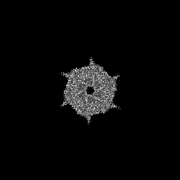
| Images | 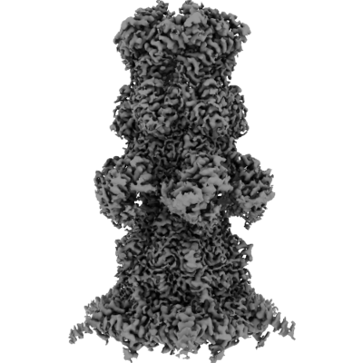 emd_22680.png emd_22680.png | 124 KB | ||
| Filedesc metadata | emd-22680.cif.gz | 7.3 KB | ||
| Archive directory |  http://ftp.pdbj.org/pub/emdb/structures/EMD-22680ftp://ftp.pdbj.org/pub/emdb/structures/EMD-22680 http://ftp.pdbj.org/pub/emdb/structures/EMD-22680ftp://ftp.pdbj.org/pub/emdb/structures/EMD-22680 | HTTPS FTP |
-Related structure data
| Related structure data | 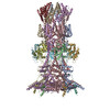 7k5cMC M: atomic model generated by this map C: citing same article ( |
|---|---|
| Similar structure data |


-Links
| EMDB pages | EMDB (EBI/PDBe) / EMDataResource |
|---|
-Map
| File | Download / File: emd_22680.map.gz / Format: CCP4 / Size: 166.4 MB / Type: IMAGE STORED AS FLOATING POINT NUMBER (4 BYTES) | ||||||||||||||||||||||||||||||||||||||||||||||||||||||||||||||||||||
|---|---|---|---|---|---|---|---|---|---|---|---|---|---|---|---|---|---|---|---|---|---|---|---|---|---|---|---|---|---|---|---|---|---|---|---|---|---|---|---|---|---|---|---|---|---|---|---|---|---|---|---|---|---|---|---|---|---|---|---|---|---|---|---|---|---|---|---|---|---|
| Annotation | Post-process map from Relion-3.0 | ||||||||||||||||||||||||||||||||||||||||||||||||||||||||||||||||||||
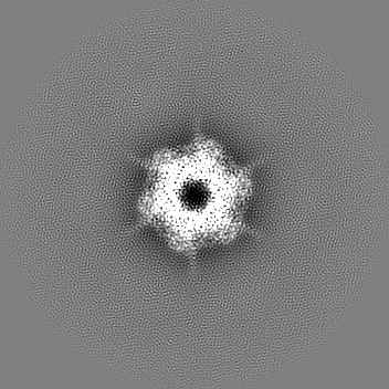
| Projections & slices | Image control
Images are generated by Spider. | ||||||||||||||||||||||||||||||||||||||||||||||||||||||||||||||||||||
| Voxel size | X=Y=Z: 1.08 Å | ||||||||||||||||||||||||||||||||||||||||||||||||||||||||||||||||||||
| Density |
| ||||||||||||||||||||||||||||||||||||||||||||||||||||||||||||||||||||
| Symmetry | Space group: 1 | ||||||||||||||||||||||||||||||||||||||||||||||||||||||||||||||||||||
| Details | EMDB XML:
CCP4 map header:
| ||||||||||||||||||||||||||||||||||||||||||||||||||||||||||||||||||||
 Z (Sec.)
 Y (Row.)
 X (Col.)


-Supplemental data
- Sample components
-Entire : Multisubunit T7 DNA ejectosome periplasmic tunnel with 6x copies ...
| Entire | Name: Multisubunit T7 DNA ejectosome periplasmic tunnel with 6x copies of Gp15 and 6x copies of Gp16 |
|---|---|
| Components |
|
-Supramolecule #1: Multisubunit T7 DNA ejectosome periplasmic tunnel with 6x copies ...
| Supramolecule | Name: Multisubunit T7 DNA ejectosome periplasmic tunnel with 6x copies of Gp15 and 6x copies of Gp16 type: complex / ID: 1 / Parent: 0 / Macromolecule list: #1-#2 |
|---|---|
| Source (natural) | Organism: Escherichia phage T7 (virus) |
| Molecular weight | Theoretical: 590 KDa |
-Macromolecule #1: Internal virion protein gp15
| Macromolecule | Name: Internal virion protein gp15 / type: protein_or_peptide / ID: 1 / Number of copies: 6 / Enantiomer: LEVO |
|---|---|
| Source (natural) | Organism: Escherichia phage T7 (virus) |
| Molecular weight | Theoretical: 84.454008 KDa |
| Recombinant expression | Organism:  |
| Sequence | String: MSKIESALQA AQPGLSRLRG GAGGMGYRAA TTQAEQPRSS LLDTIGRFAK AGADMYTAKE QRARDLADER SNEIIRKLTP EQRREALNN GTLLYQDDPY AMEALRVKTG RNAAYLVDDD VMQKIKEGVF RTREEMEEYR HSRLQEGAKV YAEQFGIDPE D VDYQRGFN ...String: MSKIESALQA AQPGLSRLRG GAGGMGYRAA TTQAEQPRSS LLDTIGRFAK AGADMYTAKE QRARDLADER SNEIIRKLTP EQRREALNN GTLLYQDDPY AMEALRVKTG RNAAYLVDDD VMQKIKEGVF RTREEMEEYR HSRLQEGAKV YAEQFGIDPE D VDYQRGFN GDITERNISL YGAHDNFLSQ QAQKGAIMNS RVELNGVLQD PDMLRRPDSA DFFEKYIDNG LVTGAIPSDA QA TQLISQA FSDASSRAGG ADFLMRVGDK KVTLNGATTT YRELIGEEQW NALMVTAQRS QFETDAKLNE QYRLKINSAL NQE DPRTAW EMLQGIKAEL DKVQPDEQMT PQREWLISAQ EQVQNQMNAW TKAQAKALDD SMKSMNKLDV IDKQFQKRIN GEWV STDFK DMPVNENTGE FKHSDMVNYA NKKLAEIDSM DIPDGAKDAM KLKYLQADSK DGAFRTAIGT MVTDAGQEWS AAVIN GKLP ERTPAMDALR RIRNADPQLI AALYPDQAEL FLTMDMMDKQ GIDPQVILDA DRLTVKRSKE QRFEDDKAFE SALNAS KAP EIARMPASLR ESARKIYDSV KYRSGNESMA MEQMTKFLKE STYTFTGDDV DGDTVGVIPK NMMQVNSDPK SWEQGRD IL EEARKGIIAS NPWITNKQLT MYSQGDSIYL MDTTGQVRVR YDKELLSKVW SENQKKLEEK AREKALADVN KRAPIVAA T KAREAAAKRV REKRKQTPKF IYGRKE UniProtKB: Internal virion protein gp15 |
-Macromolecule #2: Peptidoglycan transglycosylase gp16
| Macromolecule | Name: Peptidoglycan transglycosylase gp16 / type: protein_or_peptide / ID: 2 / Number of copies: 6 / Enantiomer: LEVO EC number: Lyases; Carbon-oxygen lyases; Acting on polysaccharides |
|---|---|
| Source (natural) | Organism: Escherichia phage T7 (virus) |
| Molecular weight | Theoretical: 144.028219 KDa |
| Recombinant expression | Organism: |
| Sequence | String: MDKYDKNVPS DYDGLFQKAA DANGVSYDLL RKVAWTESRF VPTAKSKTGP LGMMQFTKAT AKALGLRVTD GPDDDRLNPE LAINAAAKQ LAGLVGKFDG DELKAALAYN QGEGRLGNPQ LEAYSKGDFA SISEEGRNYM RNLLDVAKSP MAGQLETFGG I TPKGKGIP ...String: MDKYDKNVPS DYDGLFQKAA DANGVSYDLL RKVAWTESRF VPTAKSKTGP LGMMQFTKAT AKALGLRVTD GPDDDRLNPE LAINAAAKQ LAGLVGKFDG DELKAALAYN QGEGRLGNPQ LEAYSKGDFA SISEEGRNYM RNLLDVAKSP MAGQLETFGG I TPKGKGIP AEVGLAGIGH KQKVTQELPE STSFDVKGIE QEATAKPFAK DFWETHGETL DEYNSRSTFF GFKNAAEAEL SN SVAGMAF RAGRLDNGFD VFKDTITPTR WNSHIWTPEE LEKIRTEVKN PAYINVVTGG SPENLDDLIK LANENFENDS RAA EAGLGA KLSAGIIGAG VDPLSYVPMV GVTGKGFKLI NKALVVGAES AALNVASEGL RTSVAGGDAD YAGAALGGFV FGAG MSAIS DAVAAGLKRS KPEAEFDNEF IGPMMRLEAR ETARNANSAD LSRMNTENMK FEGEHNGVPY EDLPTERGAV VLHDG SVLS ASNPINPKTL KEFSEVDPEK AARGIKLAGF TEIGLKTLGS DDADIRRVAI DLVRSPTGMQ SGASGKFGAT ASDIHE RLH GTDQRTYNDL YKAMSDAMKD PEFSTGGAKM SREETRYTIY RRAALAIERP ELQKALTPSE RIVMDIIKRH FDTKREL ME NPAIFGNTKA VSIFPESRHK GTYVPHVYDR HAKALMIQRY GAEGLQEGIA RSWMNSYVSR PEVKARVDEM LKELHGVK E VTPEMVEKYA MDKAYGISHS DQFTNSSIIE ENIEGLVGIE NNSFLEARNL FDSDLSITMP DGQQFSVNDL RDFDMFRIM PAYDRRVNGD IAIMGSTGKT TKELKDEILA LKAKAEGDGK KTGEVHALMD TVKILTGRAR RNQDTVWETS LRAINDLGFF AKNAYMGAQ NITEIAGMIV TGNVRALGHG IPILRDTLYK SKPVSAKELK ELHASLFGKE VDQLIRPKRA DIVQRLREAT D TGPAVANI VGTLKYSTQE LAARSPWTKL LNGTTNYLLD AARQGMLGDV ISATLTGKTT RWEKEGFLRG ASVTPEQMAG IK SLIKEHM VRGEDGKFTV KDKQAFSMDP RAMDLWRLAD KVADEAMLRP HKVSLQDSHA FGALGKMVMQ FKSFTIKSLN SKF LRTFYD GYKNNRAIDA ALSIITSMGL AGGFYAMAAH VKAYALPKEK RKEYLERALD PTMIAHAALS RSSQLGAPLA MVDL VGGVL GFESSKMARS TILPKDTVKE RDPNKPYTSR EVMGAMGSNL LEQMPSAGFV ANVGATLMNA AGVVNSPNKA TEQDF MTGL MNSTKELVPN DPLTQQLVLK IYEANGVNLR ERRK UniProtKB: Peptidoglycan transglycosylase gp16 |
-Macromolecule #3: water
| Macromolecule | Name: water / type: ligand / ID: 3 / Number of copies: 219 / Formula: HOH |
|---|---|
| Molecular weight | Theoretical: 18.015 Da |
| Chemical component information |  ChemComp-HOH: |
-Experimental details
-Structure determination
| Method | cryo EM |
|---|---|
 Processing Processing | single particle reconstruction |
| Aggregation state | particle |
-Sample preparation
| Concentration | 2.2 mg/mL | ||||||||||||
|---|---|---|---|---|---|---|---|---|---|---|---|---|---|
| Buffer | pH: 8.5 Component:
Details: Solutions were made fresh and filtered. | ||||||||||||
| Grid | Model: Quantifoil R1.2/1.3 / Material: COPPER / Mesh: 300 / Support film - Material: CARBON / Support film - topology: HOLEY / Support film - Film thickness: 120 / Pretreatment - Type: GLOW DISCHARGE / Pretreatment - Time: 30 sec. / Pretreatment - Atmosphere: AIR / Pretreatment - Pressure: 0.03 kPa | ||||||||||||
| Vitrification | Cryogen name: ETHANE / Chamber humidity: 95 % / Chamber temperature: 278 K / Instrument: FEI VITROBOT MARK IV / Details: blot for 6 seconds before plunging. | ||||||||||||
| Details | This sample was monodisperse, but high concentration. Some preferred orientation. |
- Electron microscopy
| Microscope | FEI TITAN KRIOS |
|---|---|
| Image recording | Film or detector model: GATAN K3 (6k x 4k) / Number grids imaged: 1 / Number real images: 8601 / Average exposure time: 3.4 sec. / Average electron dose: 50.0 e/Å2 |
| Electron beam | Acceleration voltage: 300 kV / Electron source:  FIELD EMISSION GUN FIELD EMISSION GUN |
| Electron optics | C2 aperture diameter: 100.0 µm / Calibrated defocus max: 2.5 µm / Calibrated defocus min: 1.0 µm / Illumination mode: FLOOD BEAM / Imaging mode: BRIGHT FIELD / Cs: 2.7 mm / Nominal defocus max: 2.5 µm / Nominal defocus min: 1.0 µm / Nominal magnification: 81000 |
| Sample stage | Specimen holder model: FEI TITAN KRIOS AUTOGRID HOLDER / Cooling holder cryogen: NITROGEN |
| Experimental equipment |  Model: Titan Krios / Image courtesy: FEI Company |
+Image processing
-Atomic model buiding 1
| Refinement | Space: REAL / Protocol: AB INITIO MODEL / Overall B value: 45.1 / Target criteria: 0.92 |
|---|---|
| Output model | PDB-7k5c: |
