 Movie
Movie Controller


+ Open data

- Basic information
| Entry | Database: PDB / ID: 7k5c | ||||||
|---|---|---|---|---|---|---|---|
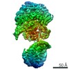
| Title | Structure of T7 DNA ejectosome periplasmic tunnel | ||||||
 Components Components |
| ||||||
 Keywords Keywords | VIRAL PROTEIN / T7 / ejectosome / ejection protein / genome-delivery / podoviridae | ||||||
| Function / homology |  Function and homology information Function and homology informationsymbiont genome ejection through host cell envelope / host cell periplasmic space / : / symbiont entry into host cell via disruption of host cell wall peptidoglycan / peptidoglycan lytic transglycosylase activity / symbiont genome ejection through host cell envelope, short tail mechanism / peptidoglycan metabolic process / symbiont entry into host cell via disruption of host cell envelope / symbiont entry into host / virion component ...symbiont genome ejection through host cell envelope / host cell periplasmic space / : / symbiont entry into host cell via disruption of host cell wall peptidoglycan / peptidoglycan lytic transglycosylase activity / symbiont genome ejection through host cell envelope, short tail mechanism / peptidoglycan metabolic process / symbiont entry into host cell via disruption of host cell envelope / symbiont entry into host / virion component / killing of cells of another organism / defense response to bacterium / hydrolase activity / host cell plasma membrane / membrane Similarity search - Function | ||||||
| Biological species |   Escherichia phage T7 (virus) Escherichia phage T7 (virus) | ||||||
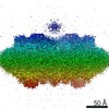
| Method | ELECTRON MICROSCOPY / single particle reconstruction / cryo EM / Resolution: 2.7 Å | ||||||
 Authors Authors | Swanson, N. / Cingolani, G. / Pumroy, R. | ||||||
| Funding support |  United States, 1items United States, 1items
| ||||||
 Citation Citation | Journal: Mol Cell / Year: 2021 Title: Cryo-EM structure of the periplasmic tunnel of T7 DNA-ejectosome at 2.7 Å resolution. Authors: Nicholas A Swanson / Ravi K Lokareddy / Fenglin Li / Chun-Feng David Hou / Sebastian Leptihn / Mikhail Pavlenok / Michael Niederweis / Ruth A Pumroy / Vera Y Moiseenkova-Bell / Gino Cingolani /  Abstract: Hershey and Chase used bacteriophage T2 genome delivery inside Escherichia coli to demonstrate that DNA, not protein, is the genetic material. Seventy years later, our understanding of viral genome ...Hershey and Chase used bacteriophage T2 genome delivery inside Escherichia coli to demonstrate that DNA, not protein, is the genetic material. Seventy years later, our understanding of viral genome delivery in prokaryotes remains limited, especially for short-tailed phages of the Podoviridae family. These viruses expel mysterious ejection proteins found inside the capsid to form a DNA-ejectosome for genome delivery into bacteria. Here, we reconstitute the phage T7 DNA-ejectosome components gp14, gp15, and gp16 and solve the periplasmic tunnel structure at 2.7 Å resolution. We find that gp14 forms an outer membrane pore, gp15 assembles into a 210 Å hexameric DNA tube spanning the host periplasm, and gp16 extends into the host cytoplasm forming a ∼4,200 residue hub. Gp16 promotes gp15 oligomerization, coordinating peptidoglycan hydrolysis, DNA binding, and lipid insertion. The reconstituted gp15:gp16 complex lacks channel-forming activity, suggesting that the pore for DNA passage forms only transiently during genome ejection. | ||||||
| History |
|
- Structure visualization
| Movie |
Movie viewer |
|---|---|
| Structure viewer | Molecule: MolmilJmol/JSmol |
- Downloads & links
-Download
| PDBx/mmCIF format | 7k5c.cif.gz | 1004.1 KB | Display | PDBx/mmCIF format |
|---|---|---|---|---|
| PDB format | pdb7k5c.ent.gz | 766.2 KB | Display | PDB format |
| PDBx/mmJSON format | 7k5c.json.gz | Tree view | PDBx/mmJSON format | |
| Others |  Other downloads Other downloads |
-Validation report
| Arichive directory | https://data.pdbj.org/pub/pdb/validation_reports/k5/7k5cftp://data.pdbj.org/pub/pdb/validation_reports/k5/7k5c | HTTPS FTP |
|---|
-Related structure data
| Related structure data | 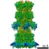 22680MC M: map data used to model this data C: citing same article ( |
|---|---|
| Similar structure data |

-Links
 PDBj
PDBj- Assembly
| Deposited unit | 
|
|---|---|
| 1 |
|
-Components
| #1: Protein | Mass: 84454.008 Da / Num. of mol.: 6 Source method: isolated from a genetically manipulated source Source: (gene. exp.) Escherichia phage T7 (virus) / Gene: 15 / Production host:  #2: Protein | Mass: 144028.219 Da / Num. of mol.: 6 Source method: isolated from a genetically manipulated source Source: (gene. exp.) Escherichia phage T7 (virus) / Gene: 16 / Production host: References: UniProt: P03726, Lyases; Carbon-oxygen lyases; Acting on polysaccharides #3: Water | ChemComp-HOH / |  Mass: 18.015 Da / Num. of mol.: 219 / Source method: isolated from a natural source / Formula: H2O Mass: 18.015 Da / Num. of mol.: 219 / Source method: isolated from a natural source / Formula: H2O |
|---|
-Experimental details
-Experiment
| Experiment | Method: ELECTRON MICROSCOPY |
|---|---|
| EM experiment | Aggregation state: PARTICLE / 3D reconstruction method: single particle reconstruction |
- Sample preparation
| Component | Name: Multisubunit T7 DNA ejectosome periplasmic tunnel with 6x copies of Gp15 and 6x copies of Gp16 Type: COMPLEX / Entity ID: #1-#2 / Source: RECOMBINANT | ||||||||||||||||||||
|---|---|---|---|---|---|---|---|---|---|---|---|---|---|---|---|---|---|---|---|---|---|
| Molecular weight | Value: 0.59 MDa | ||||||||||||||||||||
| Source (natural) | Organism: Escherichia phage T7 (virus) | ||||||||||||||||||||
| Source (recombinant) | Organism: | ||||||||||||||||||||
| Buffer solution | pH: 8.5 / Details: Solutions were made fresh and filtered. | ||||||||||||||||||||
| Buffer component |
| ||||||||||||||||||||
| Specimen | Conc.: 2.2 mg/ml / Embedding applied: NO / Shadowing applied: NO / Staining applied: NO / Vitrification applied: YES Details: This sample was monodisperse, but high concentration. Some preferred orientation. | ||||||||||||||||||||
| Specimen support | Grid material: COPPER / Grid mesh size: 300 divisions/in. / Grid type: Quantifoil R1.2/1.3 | ||||||||||||||||||||
| Vitrification | Instrument: FEI VITROBOT MARK IV / Cryogen name: ETHANE / Humidity: 95 % / Chamber temperature: 278 K / Details: blot for 6 seconds before plunging |
- Electron microscopy imaging
| Experimental equipment |  Model: Titan Krios / Image courtesy: FEI Company |
|---|---|
| Microscopy | Model: FEI TITAN KRIOS |
| Electron gun | Electron source:  FIELD EMISSION GUN / Accelerating voltage: 300 kV / Illumination mode: FLOOD BEAM FIELD EMISSION GUN / Accelerating voltage: 300 kV / Illumination mode: FLOOD BEAM |
| Electron lens | Mode: BRIGHT FIELD / Nominal magnification: 81000 X / Nominal defocus max: 2500 nm / Nominal defocus min: 1000 nm / Calibrated defocus min: 1000 nm / Calibrated defocus max: 2500 nm / Cs: 2.7 mm / C2 aperture diameter: 100 µm |
| Specimen holder | Cryogen: NITROGEN / Specimen holder model: FEI TITAN KRIOS AUTOGRID HOLDER |
| Image recording | Average exposure time: 3.4 sec. / Electron dose: 50 e/Å2 / Film or detector model: GATAN K3 (6k x 4k) / Num. of grids imaged: 1 / Num. of real images: 8601 |
- Processing
| EM software |
| ||||||||||||||||||||||||||||||||||||||||||||||||||
|---|---|---|---|---|---|---|---|---|---|---|---|---|---|---|---|---|---|---|---|---|---|---|---|---|---|---|---|---|---|---|---|---|---|---|---|---|---|---|---|---|---|---|---|---|---|---|---|---|---|---|---|
| CTF correction | Type: PHASE FLIPPING AND AMPLITUDE CORRECTION | ||||||||||||||||||||||||||||||||||||||||||||||||||
| Particle selection | Num. of particles selected: 4700000 Details: Manually picked 2000 particles using Relion GUI, followed by 2D-Classification to select templates, then Relion auto-picking was used. | ||||||||||||||||||||||||||||||||||||||||||||||||||
| Symmetry | Point symmetry: C1 (asymmetric) | ||||||||||||||||||||||||||||||||||||||||||||||||||
| 3D reconstruction | Resolution: 2.7 Å / Resolution method: FSC 0.143 CUT-OFF / Num. of particles: 208024 / Num. of class averages: 2 / Symmetry type: POINT | ||||||||||||||||||||||||||||||||||||||||||||||||||
| Atomic model building | B value: 45.1 / Protocol: AB INITIO MODEL / Space: REAL / Target criteria: 0.92 |
