Movie
Movie Controller

[English] 日本語
 Yorodumi
- EMDB-0722: Full length S-OPA1 coated liposome tube at nucleotide-free state -
+ Open data

- Basic information
| Entry | Database: EMDB / ID: EMD-0722 | ||||||||||||
|---|---|---|---|---|---|---|---|---|---|---|---|---|---|
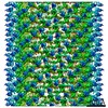
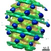
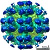
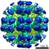
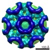
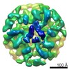
| Title | Full length S-OPA1 coated liposome tube at nucleotide-free state | ||||||||||||
 Map data Map data | |||||||||||||
 Sample Sample |
| ||||||||||||
| Biological species |  Homo sapiens (human) Homo sapiens (human) | ||||||||||||
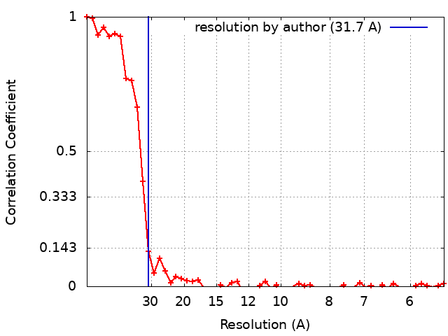
| Method | subtomogram averaging / cryo EM / Resolution: 31.7 Å | ||||||||||||
 Authors Authors | Zhang D / Niu T / Zhang Y / Sun F | ||||||||||||
| Funding support |  China, 3 items China, 3 items
| ||||||||||||
 Citation Citation | Journal: Elife / Year: 2020 Title: Cryo-EM structures of S-OPA1 reveal its interactions with membrane and changes upon nucleotide binding. Authors: Danyang Zhang / Yan Zhang / Jun Ma / Chunmei Zhu / Tongxin Niu / Wenbo Chen / Xiaoyun Pang / Yujia Zhai / Fei Sun / Abstract: Mammalian mitochondrial inner membrane fusion is mediated by optic atrophy 1 (OPA1). Under physiological conditions, OPA1 undergoes proteolytic processing to form a membrane-anchored long isoform (L- ...Mammalian mitochondrial inner membrane fusion is mediated by optic atrophy 1 (OPA1). Under physiological conditions, OPA1 undergoes proteolytic processing to form a membrane-anchored long isoform (L-OPA1) and a soluble short isoform (S-OPA1). A combination of L-OPA1 and S-OPA1 is essential for efficient membrane fusion; however, the relevant mechanism is not well understood. In this study, we investigate the cryo-electron microscopic structures of S-OPA1-coated liposomes in nucleotide-free and GTPγS-bound states. S-OPA1 exhibits a general dynamin-like structure and can assemble onto membranes in a helical array with a dimer building block. We reveal that hydrophobic residues in its extended membrane-binding domain are critical for its tubulation activity. The binding of GTPγS triggers a conformational change and results in a rearrangement of the helical lattice and tube expansion similar to that of S-Mgm1. These observations indicate that S-OPA1 adopts a dynamin-like power stroke membrane remodeling mechanism during mitochondrial inner membrane fusion. | ||||||||||||
| History |
|
- Structure visualization
| Movie |
 Movie viewer Movie viewer |
|---|---|
| Structure viewer | EM map: SurfViewMolmilJmol/JSmol |
| Supplemental images |

- Downloads & links
-EMDB archive
| Map data | emd_0722.map.gz | 6 MB | EMDB map data format | |
|---|---|---|---|---|
| Header (meta data) | emd-0722-v30.xmlemd-0722.xml | 14.4 KB 14.4 KB | Display Display | EMDB header |
| FSC (resolution estimation) | emd_0722_fsc.xml | 5.5 KB | Display | FSC data file |
| Images | 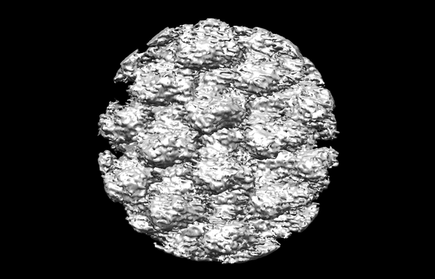 emd_0722.png emd_0722.png | 91.6 KB | ||
| Archive directory |  http://ftp.pdbj.org/pub/emdb/structures/EMD-0722ftp://ftp.pdbj.org/pub/emdb/structures/EMD-0722 http://ftp.pdbj.org/pub/emdb/structures/EMD-0722ftp://ftp.pdbj.org/pub/emdb/structures/EMD-0722 | HTTPS FTP |
-Related structure data


-Links
| EMDB pages | EMDB (EBI/PDBe) / EMDataResource |
|---|
-Map
| File | Download / File: emd_0722.map.gz / Format: CCP4 / Size: 8 MB / Type: IMAGE STORED AS FLOATING POINT NUMBER (4 BYTES) | ||||||||||||||||||||||||||||||||||||||||||||||||||||||||||||
|---|---|---|---|---|---|---|---|---|---|---|---|---|---|---|---|---|---|---|---|---|---|---|---|---|---|---|---|---|---|---|---|---|---|---|---|---|---|---|---|---|---|---|---|---|---|---|---|---|---|---|---|---|---|---|---|---|---|---|---|---|---|
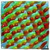
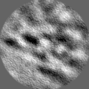
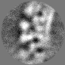
| Projections & slices | Image control
Images are generated by Spider. | ||||||||||||||||||||||||||||||||||||||||||||||||||||||||||||
| Voxel size | X=Y=Z: 2.72 Å | ||||||||||||||||||||||||||||||||||||||||||||||||||||||||||||
| Density |
| ||||||||||||||||||||||||||||||||||||||||||||||||||||||||||||
| Symmetry | Space group: 1 | ||||||||||||||||||||||||||||||||||||||||||||||||||||||||||||
| Details | EMDB XML:
CCP4 map header:
| ||||||||||||||||||||||||||||||||||||||||||||||||||||||||||||
 Z (Sec.)
 Y (Row.)
 X (Col.)


-Supplemental data
- Sample components
-Entire : full length S-OPA1 coated liposome tube
| Entire | Name: full length S-OPA1 coated liposome tube |
|---|---|
| Components |
|
-Supramolecule #1: full length S-OPA1 coated liposome tube
| Supramolecule | Name: full length S-OPA1 coated liposome tube / type: complex / ID: 1 / Parent: 0 / Macromolecule list: all Details: 1mg/ml full length S-OPA1 was incubated with 1mg/ml liposome at room temperature for 30 min. |
|---|---|
| Source (natural) | Organism: Homo sapiens (human) |
| Recombinant expression | Organism:  |
-Macromolecule #1: Dynamin-like 120 kDa protein, mitochondrial, short form for isofo...
| Macromolecule | Name: Dynamin-like 120 kDa protein, mitochondrial, short form for isoform 1, Optic atrophy protein 1 (OPA1), short form for isoform 1 type: protein_or_peptide / ID: 1 / Enantiomer: LEVO |
|---|---|
| Source (natural) | Organism: Homo sapiens (human) |
| Recombinant expression | Organism: |
| Sequence | String: GPGSTDRGSE SDKHFRKVSD KEKIDQLQEE LLHTQLKYQR ILERLEKEN KELRKLVLQK DDKGIHHRKL KKSLIDMYSE VLDVLSDYDA SYNTQDHLPR VVVVGDQSAG KTSVLEMIA QARIFPRGSG EMMTRSPVKV TLSEGPHHVA LFKDSSREFD LTKEEDLAAL R ...String: GPGSTDRGSE SDKHFRKVSD KEKIDQLQEE LLHTQLKYQR ILERLEKEN KELRKLVLQK DDKGIHHRKL KKSLIDMYSE VLDVLSDYDA SYNTQDHLPR VVVVGDQSAG KTSVLEMIA QARIFPRGSG EMMTRSPVKV TLSEGPHHVA LFKDSSREFD LTKEEDLAAL R HEIELRMR KNVKEGCTVS PETISLNVKG PGLQRMVLVD LPGVINTVTS GMAPDTKETI FS ISKAYMQ NPNAIILCIQ DGSVDAERSI VTDLVSQMDP HGRRTIFVLT KVDLAEKNVA SPS RIQQII EGKLFPMKAL GYFAVVTGKG NSSESIEAIR EYEEEFFQNS KLLKTSMLKA HQVT TRNLS LAVSDCFWKM VRESVEQQAD SFKATRFNLE TEWKNNYPRL RELDRNELFE KAKNE ILDE VISLSQVTPK HWEEILQQSL WERVSTHVIE NIYLPAAQTM NSGTFNTTVD IKLKQW TDK QLPNKAVEVA WETLQEEFSR FMTEPKGKEH DDIFDKLKEA VKEESIKRHK WNDFAED SL RVIQHNALED RSISDKQQWD AAIYFMEEAL QARLKDTENA IENMVGPDWK KRWLYWKN R TQEQCVHNET KNELEKMLKC NEEHPAYLAS DEITTVRKNL ESRGVEVDPS LIKDTWHQV YRRHFLKTAL NHCNLCRRGF YYYQRHFVDS ELECNDVVLF WRIQRMLAIT ANTLRQQLTN TEVRRLEKN VKEVLEDFAE DGEKKIKLLT GKRVQLAEDL KKVREIQEKL DAFIEALHQE K |
-Experimental details
-Structure determination
| Method | cryo EM |
|---|---|
 Processing Processing | subtomogram averaging |
| Aggregation state | helical array |
-Sample preparation
| Concentration | 1 mg/mL | ||||||||
|---|---|---|---|---|---|---|---|---|---|
| Buffer | pH: 8 Component:
| ||||||||
| Grid | Model: Quantifoil R2/1 / Material: COPPER / Mesh: 300 / Support film - Material: CARBON / Pretreatment - Type: GLOW DISCHARGE / Pretreatment - Atmosphere: OTHER | ||||||||
| Vitrification | Cryogen name: ETHANE / Chamber humidity: 100 % / Chamber temperature: 289 K / Instrument: FEI VITROBOT MARK IV Details: The grid was blotted for 3.5s with force 1 before plunging.. | ||||||||
| Details | 1mg/ml full length S-OPA1 was incubated with 1mg/ml liposome at room temperature for 30 min. |
- Electron microscopy
| Microscope | FEI TITAN KRIOS |
|---|---|
| Image recording | Film or detector model: GATAN K2 SUMMIT (4k x 4k) / Detector mode: COUNTING / Digitization - Dimensions - Width: 3838 pixel / Digitization - Dimensions - Height: 3710 pixel / Digitization - Frames/image: 1-12 / Number grids imaged: 1 / Average exposure time: 1.0 sec. / Average electron dose: 3.0 e/Å2 |
| Electron beam | Acceleration voltage: 300 kV / Electron source:  FIELD EMISSION GUN FIELD EMISSION GUN |
| Electron optics | Illumination mode: FLOOD BEAM / Imaging mode: BRIGHT FIELD / Cs: 2.7 mm / Nominal defocus max: 5.0 µm / Nominal defocus min: 4.0 µm |
| Sample stage | Specimen holder model: FEI TITAN KRIOS AUTOGRID HOLDER / Cooling holder cryogen: NITROGEN |
| Experimental equipment |  Model: Titan Krios / Image courtesy: FEI Company |
+Image processing
-Atomic model buiding 1
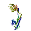
| Initial model | PDB ID: Chain - Chain ID: A |
|---|---|
| Refinement | Space: REAL / Protocol: RIGID BODY FIT |